1QBN

Bovine Trypsin 2-[amino(imino)methyl]-2-hydroxyphenoxy]-6-[3-(4,5-dihydro-1H-imidazol-2-yl)phenoxy]pyridine-4-carboxylic Acid (ZK-806688) Complex


Experimental Data Snapshot

  • Method: X-RAY DIFFRACTION
  • Resolution: 1.80 Å
  • R-Value Free: 0.217 
  • R-Value Work: 0.155 

wwPDB Validation   3D Report Full Report


This is version 1.2 of the entry. See complete history


Literature

Crystallographic analysis of potent and selective factor Xa inhibitors complexed to bovine trypsin.

Whitlow, M.Arnaiz, D.O.Buckman, B.O.Davey, D.D.Griedel, B.Guilford, W.J.Koovakkat, S.K.Liang, A.Mohan, R.Phillips, G.B.Seto, M.Shaw, K.J.Xu, W.Zhao, Z.Light, D.R.Morrissey, M.M.

(1999) Acta Crystallogr D Biol Crystallogr 55: 1395-1404

  • DOI: https://doi.org/10.1107/s0907444999007350
  • Primary Citation of Related Structures:  
    1QA0, 1QB1, 1QB6, 1QB9, 1QBN, 1QBO

  • PubMed Abstract: 

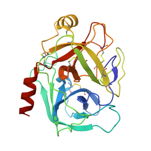
    Factor Xa is a serine protease which activates thrombin (factor IIa) and plays a key regulatory role in the blood-coagulation cascade. Factor Xa is, therefore, an important target for the design of anti-thrombotics. Both factor Xa and thrombin share sequence and structural homology with trypsin. As part of a factor Xa inhibitor-design program, a number of factor Xa inhibitors were crystallographically studied complexed to bovine trypsin. The structures of one diaryl benzimidazole, one diaryl carbazole and three diaryloxypyridines are described. All five compounds bind to trypsin in an extended conformation, with an amidinoaryl group in the S1 pocket and a second basic/hydrophobic moiety bound in the S4 pocket. These binding modes all bear a resemblance to the reported binding mode of DX-9065a in bovine trypsin and human factor Xa.


  • Organizational Affiliation

    Berlex Biosciences, 15049 San Pablo Avenue, PO Box 4099, Richmond, California 94804, USA. marc_whitlow@berlex.com


Macromolecules
Find similar proteins by:  (by identity cutoff)  |  3D Structure
Entity ID: 1
MoleculeChains Sequence LengthOrganismDetailsImage
PROTEIN (TRYPSIN)223Bos taurusMutation(s): 0 
EC: 3.4.21.4
UniProt
Find proteins for P00760 (Bos taurus)
Explore P00760 
Go to UniProtKB:  P00760
Entity Groups  
Sequence Clusters30% Identity50% Identity70% Identity90% Identity95% Identity100% Identity
UniProt GroupP00760
Sequence Annotations
Expand
  • Reference Sequence
Small Molecules
Ligands 2 Unique
IDChains Name / Formula / InChI Key2D Diagram3D Interactions
688
Query on 688

Download Ideal Coordinates CCD File 
D [auth A]2-[AMINO(IMINO)METHYL]-2-HYDROXYPHENOXY]-6-[3-(4,5-DIHYDRO-1H-IMIDAZOL-2-YL)PHENOXY]PYRIDINE-4-CARBOXYLIC ACID
C22 H17 N5 O5
ILFWHJBPGCBXAY-UHFFFAOYSA-N
CA
Query on CA

Download Ideal Coordinates CCD File 
B [auth A],
C [auth A]
CALCIUM ION
Ca
BHPQYMZQTOCNFJ-UHFFFAOYSA-N
Binding Affinity Annotations 
IDSourceBinding Affinity
688 Binding MOAD:  1QBN Ki: 1400 (nM) from 1 assay(s)
PDBBind:  1QBN Ki: 1400 (nM) from 1 assay(s)
Experimental Data & Validation

Experimental Data

  • Method: X-RAY DIFFRACTION
  • Resolution: 1.80 Å
  • R-Value Free: 0.217 
  • R-Value Work: 0.155 
  • Space Group: P 31 2 1
Unit Cell:
Length ( Å )Angle ( ˚ )
a = 55.02α = 90
b = 55.02β = 90
c = 109.14γ = 120
Software Package:
Software NamePurpose
DENZOdata reduction
SCALEPACKdata scaling
PROFFTrefinement

Structure Validation

View Full Validation Report


Entry History 

Deposition Data

  • Released Date: 2000-05-03 
  • Deposition Author(s): Whitlow, M.

Revision History  (Full details and data files)

  • Version 1.0: 2000-05-03
    Type: Initial release
  • Version 1.1: 2007-10-21
    Changes: Version format compliance
  • Version 1.2: 2011-07-13
    Changes: Version format compliance