1QBK

STRUCTURE OF THE KARYOPHERIN BETA2-RAN GPPNHP NUCLEAR TRANSPORT COMPLEX


Experimental Data Snapshot

  • Method: X-RAY DIFFRACTION
  • Resolution: 3.00 Å
  • R-Value Free: 0.315 
  • R-Value Work: 0.267 
  • R-Value Observed: 0.267 

wwPDB Validation   3D Report Full Report


This is version 1.4 of the entry. See complete history


Literature

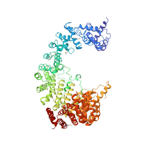
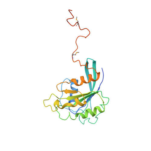
Structure of the nuclear transport complex karyopherin-beta2-Ran x GppNHp.

Chook, Y.M.Blobel, G.

(1999) Nature 399: 230-237

  • DOI: https://doi.org/10.1038/20375
  • Primary Citation of Related Structures:  
    1QBK

  • PubMed Abstract: 

    Transport factors in the karyopherin-beta (also called importin-beta) family mediate the movement of macromolecules in nuclear-cytoplasmic transport pathways. Karyopherin-beta2 (transportin) binds a cognate import substrate and targets it to the nuclear pore complex. In the nucleus, Ran x GTP binds karyopherin-beta2 and dissociates the substrate. Here we present the 3.0 A structure of the karyopherin-beta2-Ran x GppNHp complex where GppNHp is a non-hydrolysable GTP analogue. Karyopherin-beta2 contains eighteen HEAT repeats arranged into two continuous orthogonal arches. Ran is clamped in the amino-terminal arch and substrate-binding activity is mapped to the carboxy-terminal arch. A large loop in HEAT repeat 7 spans both arches. Interactions of the loop with Ran and the C-terminal arch implicate it in GTPase-mediated dissociation of the import-substrate. Ran x GppNHp in the complex shows extensive structural rearrangement, compared to Ran GDP, in regions contacting karyopherin-beta2. This provides a structural basis for the specificity of the karyopherin-beta family for the GTP-bound state of Ran, as well as a rationale for interactions of the karyopherin-Ran complex with the regulatory proteins ranGAP, ranGEF and ranBP1.


  • Organizational Affiliation

    Laboratory of Cell Biology, Howard Hughes Medical Institute, The Rockefeller University, New York, New York 10021, USA.


Macromolecules
Find similar proteins by:  (by identity cutoff)  |  3D Structure
Entity ID: 1
MoleculeChains Sequence LengthOrganismDetailsImage
KARYOPHERIN BETA2A [auth B]890Homo sapiensMutation(s): 24 
UniProt & NIH Common Fund Data Resources
Find proteins for Q92973 (Homo sapiens)
Explore Q92973 
Go to UniProtKB:  Q92973
PHAROS:  Q92973
GTEx:  ENSG00000083312 
Entity Groups  
Sequence Clusters30% Identity50% Identity70% Identity90% Identity95% Identity100% Identity
UniProt GroupQ92973
Sequence Annotations
Expand
  • Reference Sequence
Find similar proteins by:  (by identity cutoff)  |  3D Structure
Entity ID: 2
MoleculeChains Sequence LengthOrganismDetailsImage
RANB [auth C]216Homo sapiensMutation(s): 3 
UniProt & NIH Common Fund Data Resources
Find proteins for P62826 (Homo sapiens)
Explore P62826 
Go to UniProtKB:  P62826
PHAROS:  P62826
GTEx:  ENSG00000132341 
Entity Groups  
Sequence Clusters30% Identity50% Identity70% Identity90% Identity95% Identity100% Identity
UniProt GroupP62826
Sequence Annotations
Expand
  • Reference Sequence
Small Molecules
Ligands 2 Unique
IDChains Name / Formula / InChI Key2D Diagram3D Interactions
GNP
Query on GNP

Download Ideal Coordinates CCD File 
D [auth C]PHOSPHOAMINOPHOSPHONIC ACID-GUANYLATE ESTER
C10 H17 N6 O13 P3
UQABYHGXWYXDTK-UUOKFMHZSA-N
MG
Query on MG

Download Ideal Coordinates CCD File 
C
MAGNESIUM ION
Mg
JLVVSXFLKOJNIY-UHFFFAOYSA-N
Modified Residues  1 Unique
IDChains TypeFormula2D DiagramParent
MSE
Query on MSE
A [auth B]L-PEPTIDE LINKINGC5 H11 N O2 SeMET
Experimental Data & Validation

Experimental Data

  • Method: X-RAY DIFFRACTION
  • Resolution: 3.00 Å
  • R-Value Free: 0.315 
  • R-Value Work: 0.267 
  • R-Value Observed: 0.267 
  • Space Group: P 32 2 1
Unit Cell:
Length ( Å )Angle ( ˚ )
a = 133.11α = 90
b = 133.11β = 90
c = 138.41γ = 120
Software Package:
Software NamePurpose
MLPHAREphasing
CNSrefinement
DENZOdata reduction
SCALEPACKdata scaling

Structure Validation

View Full Validation Report


Entry History 

Deposition Data

Revision History  (Full details and data files)

  • Version 1.0: 1999-06-01
    Type: Initial release
  • Version 1.1: 2008-04-27
    Changes: Version format compliance
  • Version 1.2: 2011-07-13
    Changes: Version format compliance
  • Version 1.3: 2018-01-31
    Changes: Experimental preparation
  • Version 1.4: 2024-03-13
    Changes: Data collection, Database references, Derived calculations, Source and taxonomy, Structure summary