1QBI

SOLUBLE QUINOPROTEIN GLUCOSE DEHYDROGENASE FROM ACINETOBACTER CALCOACETICUS


Experimental Data Snapshot

  • Method: X-RAY DIFFRACTION
  • Resolution: 1.72 Å
  • R-Value Free: 0.208 
  • R-Value Work: 0.165 
  • R-Value Observed: 0.166 

wwPDB Validation   3D Report Full Report


This is version 1.5 of the entry. See complete history


Literature

The 1.7 A crystal structure of the apo form of the soluble quinoprotein glucose dehydrogenase from Acinetobacter calcoaceticus reveals a novel internal conserved sequence repeat.

Oubrie, A.Rozeboom, H.J.Kalk, K.H.Duine, J.A.Dijkstra, B.W.

(1999) J Mol Biol 289: 319-333

  • DOI: https://doi.org/10.1006/jmbi.1999.2766
  • Primary Citation of Related Structures:  
    1QBI

  • PubMed Abstract: 

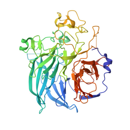
    The crystal structure of a dimeric apo form of the soluble quinoprotein glucose dehydrogenase (s-GDH) from Acinetobacter calcoaceticus has been solved by multiple isomorphous replacement followed by density modification, and was subsequently refined at 1. 72 A resolution to a final crystallographic R-factor of 16.5% and free R-factor of 20.8% [corrected]. The s-GDH monomer has a beta-propeller fold consisting of six four-stranded anti-parallel beta-sheets aligned around a pseudo 6-fold symmetry axis. The enzyme binds three calcium ions per monomer, two of which are located in the dimer interface. The third is bound in the putative active site, where it may bind and functionalize the pyrroloquinoline quinone (PQQ) cofactor. A data base search unexpectedly showed that four uncharacterized protein sequences are homologous to s-GDH with many residues in the putative active site absolutely conserved. This indicates that these homologs may have a similar structure and that they may catalyze similar PQQ-dependent reactions.A structure-based sequence alignment of the six four-stranded beta-sheets in s-GDH's beta-propeller fold shows an internally conserved sequence repeat that gives rise to two distinct conserved structural motifs. The first structural motif is found at the corner of the short beta-turn between the inner two beta-strands of the beta-sheets, where an Asp side-chain points back into the beta-sheet to form a hydrogen-bond with the OH/NH of a Tyr/Trp side-chain in the same beta-sheet. The second motif involves an Arg/Lys side-chain in the C beta-strand of one beta-sheet, which forms a bidentate salt-bridge with an Asp/Glu in the CD loop of the next beta-sheet. These intra and inter-beta-sheet hydrogen-bonds are likely to contribute to the stability of the s-GDH beta-propeller fold.


  • Organizational Affiliation

    Laboratory of Biophysical Chemistry and BIOSON Research Institute, University of Groningen, Nijenborgh 4, Groningen, 9747 AG, The Netherlands.


Macromolecules
Find similar proteins by:  (by identity cutoff)  |  3D Structure
Entity ID: 1
MoleculeChains Sequence LengthOrganismDetailsImage
SOLUBLE QUINOPROTEIN GLUCOSE DEHYDROGENASE
A, B
454Acinetobacter calcoaceticusMutation(s): 0 
UniProt
Find proteins for P13650 (Acinetobacter calcoaceticus)
Explore P13650 
Go to UniProtKB:  P13650
Entity Groups  
Sequence Clusters30% Identity50% Identity70% Identity90% Identity95% Identity100% Identity
UniProt GroupP13650
Sequence Annotations
Expand
  • Reference Sequence
Small Molecules
Ligands 3 Unique
IDChains Name / Formula / InChI Key2D Diagram3D Interactions
PT
Query on PT

Download Ideal Coordinates CCD File 
C [auth A],
H [auth B]
PLATINUM (II) ION
Pt
HRGDZIGMBDGFTC-UHFFFAOYSA-N
GOL
Query on GOL

Download Ideal Coordinates CCD File 
G [auth A]GLYCEROL
C3 H8 O3
PEDCQBHIVMGVHV-UHFFFAOYSA-N
CA
Query on CA

Download Ideal Coordinates CCD File 
D [auth A]
E [auth A]
F [auth A]
I [auth B]
J [auth B]
D [auth A],
E [auth A],
F [auth A],
I [auth B],
J [auth B],
K [auth B]
CALCIUM ION
Ca
BHPQYMZQTOCNFJ-UHFFFAOYSA-N
Experimental Data & Validation

Experimental Data

  • Method: X-RAY DIFFRACTION
  • Resolution: 1.72 Å
  • R-Value Free: 0.208 
  • R-Value Work: 0.165 
  • R-Value Observed: 0.166 
  • Space Group: P 1 21 1
Unit Cell:
Length ( Å )Angle ( ˚ )
a = 60.148α = 90
b = 92.1β = 105.17
c = 84.91γ = 90
Software Package:
Software NamePurpose
X-PLORrefinement
SCALEPACKdata scaling
PHASESphasing
REFMACrefinement
DENZOdata reduction

Structure Validation

View Full Validation Report


Entry History 

Deposition Data

Revision History  (Full details and data files)

  • Version 1.0: 2000-02-25
    Type: Initial release
  • Version 1.1: 2007-10-16
    Changes: Version format compliance
  • Version 1.2: 2011-07-13
    Changes: Non-polymer description, Version format compliance
  • Version 1.3: 2019-07-24
    Changes: Data collection, Refinement description
  • Version 1.4: 2019-08-14
    Changes: Data collection
  • Version 1.5: 2024-02-14
    Changes: Data collection, Database references, Derived calculations