1QB4

CRYSTAL STRUCTURE OF MN(2+)-BOUND PHOSPHOENOLPYRUVATE CARBOXYLASE


Experimental Data Snapshot

  • Method: X-RAY DIFFRACTION
  • Resolution: 2.60 Å
  • R-Value Free: 0.261 
  • R-Value Work: 0.223 
  • R-Value Observed: 0.221 

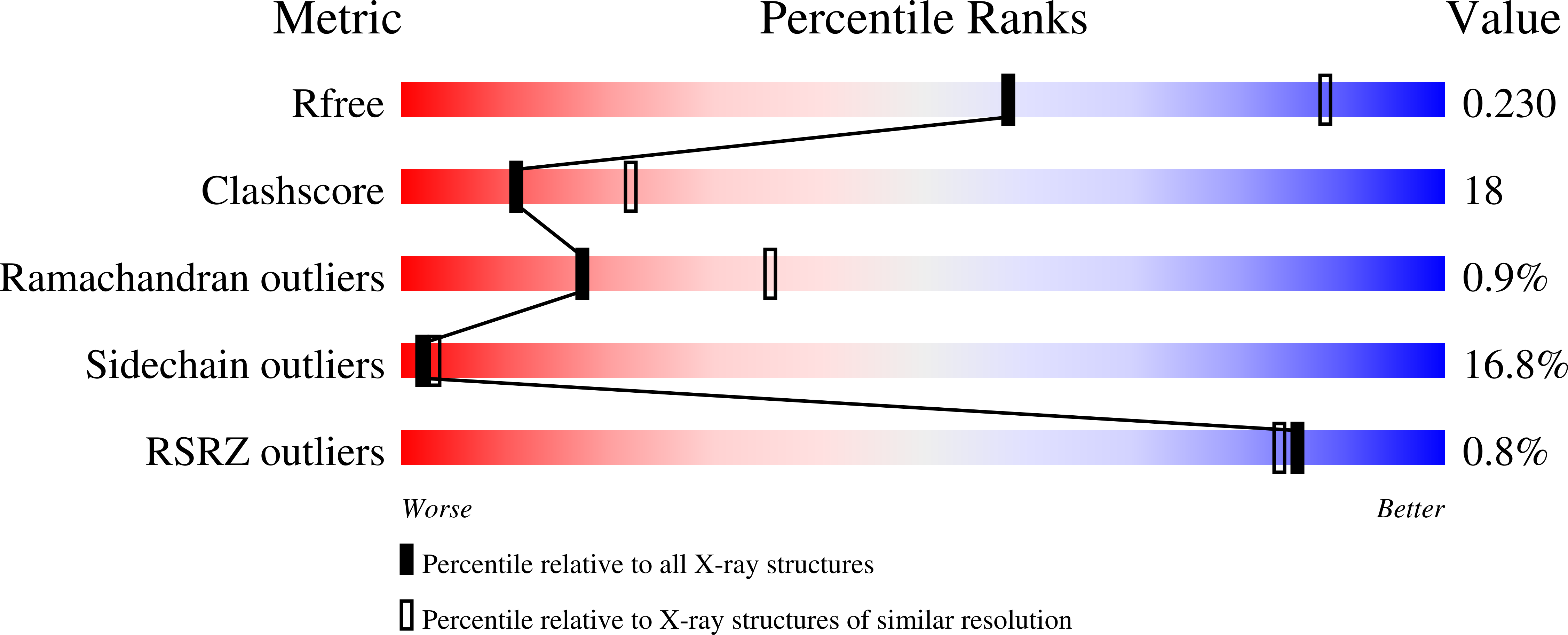
wwPDB Validation   3D Report Full Report


This is version 1.4 of the entry. See complete history


Literature

Plausible phosphoenolpyruvate binding site revealed by 2.6 A structure of Mn2+-bound phosphoenolpyruvate carboxylase from Escherichia coli

Matsumura, H.Terada, M.Shirakata, S.Inoue, T.Yoshinaga, T.Izui, K.Kai, Y.

(1999) FEBS Lett 458: 93-96

  • DOI: https://doi.org/10.1016/s0014-5793(99)01103-5
  • Primary Citation of Related Structures:  
    1QB4

  • PubMed Abstract: 

    We have determined the crystal structure of Mn2+-bound Escherichia coli phosphoenolpyruvate carboxylase (PEPC) using X-ray diffraction at 2.6 A resolution, and specified the location of enzyme-bound Mn2+, which is essential for catalytic activity. The electron density map reveals that Mn2+ is bound to the side chain oxygens of Glu-506 and Asp-543, and located at the top of the alpha/beta barrel in PEPC. The coordination sphere of Mn2+ observed in E. coli PEPC is similar to that of Mn2+ found in the pyruvate kinase structure. The model study of Mn2+-bound PEPC complexed with phosphoenolpyruvate (PEP) reveals that the side chains of Arg-396, Arg-581 and Arg-713 could interact with PEP.


  • Organizational Affiliation

    Department of Materials Chemistry, Graduate School of Engineering, Osaka University, Japan.


Macromolecules
Find similar proteins by:  (by identity cutoff)  |  3D Structure
Entity ID: 1
MoleculeChains Sequence LengthOrganismDetailsImage
PHOSPHOENOLPYRUVATE CARBOXYLASE883Escherichia coliMutation(s): 0 
Gene Names: K12
EC: 4.1.1.31
UniProt
Find proteins for P00864 (Escherichia coli (strain K12))
Explore P00864 
Go to UniProtKB:  P00864
Entity Groups  
Sequence Clusters30% Identity50% Identity70% Identity90% Identity95% Identity100% Identity
UniProt GroupP00864
Sequence Annotations
Expand
  • Reference Sequence
Small Molecules
Experimental Data & Validation

Experimental Data

  • Method: X-RAY DIFFRACTION
  • Resolution: 2.60 Å
  • R-Value Free: 0.261 
  • R-Value Work: 0.223 
  • R-Value Observed: 0.221 
  • Space Group: I 2 2 2
Unit Cell:
Length ( Å )Angle ( ˚ )
a = 117.75α = 90
b = 248.41β = 90
c = 83.47γ = 90
Software Package:
Software NamePurpose
AMoREphasing
REFMACrefinement
DENZOdata reduction
SCALEPACKdata scaling

Structure Validation

View Full Validation Report


Entry History 

Deposition Data

Revision History  (Full details and data files)

  • Version 1.0: 2002-05-01
    Type: Initial release
  • Version 1.1: 2008-04-27
    Changes: Version format compliance
  • Version 1.2: 2011-07-13
    Changes: Version format compliance
  • Version 1.3: 2018-05-23
    Changes: Data collection, Derived calculations
  • Version 1.4: 2024-02-14
    Changes: Data collection, Database references, Derived calculations