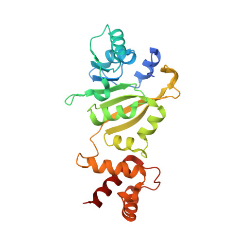
1QAQ

THE STRUCTURE OF THE RRNA METHYLTRANSFERASE ERMC': IMPLICATIONS FOR THE REACTION MECHANISM


Experimental Data Snapshot

  • Method: X-RAY DIFFRACTION
  • Resolution: 2.80 Å
  • R-Value Free: 0.284 
  • R-Value Work: 0.229 

wwPDB Validation   3D Report Full Report


This is version 1.5 of the entry. See complete history


Literature

The 2.2 A structure of the rRNA methyltransferase ErmC' and its complexes with cofactor and cofactor analogs: implications for the reaction mechanism.

Schluckebier, G.Zhong, P.Stewart, K.D.Kavanaugh, T.J.Abad-Zapatero, C.

(1999) J Mol Biol 289: 277-291

  • DOI: https://doi.org/10.1006/jmbi.1999.2788
  • Primary Citation of Related Structures:  
    1QAM, 1QAN, 1QAO, 1QAQ

  • PubMed Abstract: 

    The rRNA methyltransferase ErmC' transfers methyl groups from S -adenosyl-l-methionine to atom N6 of an adenine base within the peptidyltransferase loop of 23 S rRNA, thus conferring antibiotic resistance against a number of macrolide antibiotics. The crystal structures of ErmC' and of its complexes with the cofactor S -adenosyl-l-methionine, the reaction product S-adenosyl-l-homocysteine and the methyltransferase inhibitor Sinefungin, respectively, show that the enzyme undergoes small conformational changes upon ligand binding. Overall, the ligand molecules bind to the protein in a similar mode as observed for other methyltransferases. Small differences between the binding of the amino acid parts of the different ligands are correlated with differences in their chemical structure. A model for the transition-state based on the atomic details of the active site is consistent with a one-step methyl-transfer mechanism and might serve as a first step towards the design of potent Erm inhibitors.


  • Organizational Affiliation

    Abbott Laboratories, D46Y-AP 10, 100 Abbott Park Road, Abbott Park, IL, 60064, USA. gerd.schluckebier@abbott.com


Macromolecules
Find similar proteins by:  (by identity cutoff)  |  3D Structure
Entity ID: 1
MoleculeChains Sequence LengthOrganismDetailsImage
ERMC' RRNA METHYLTRANSFERASE244Bacillus subtilisMutation(s): 0 
EC: 2.1.1.48
UniProt
Find proteins for P13956 (Bacillus subtilis)
Explore P13956 
Go to UniProtKB:  P13956
Entity Groups  
Sequence Clusters30% Identity50% Identity70% Identity90% Identity95% Identity100% Identity
UniProt GroupP13956
Sequence Annotations
Expand
  • Reference Sequence
Small Molecules
Ligands 1 Unique
IDChains Name / Formula / InChI Key2D Diagram3D Interactions
SFG
Query on SFG

Download Ideal Coordinates CCD File 
B [auth A]SINEFUNGIN
C15 H23 N7 O5
LMXOHSDXUQEUSF-YECHIGJVSA-N
Binding Affinity Annotations 
IDSourceBinding Affinity
SFG PDBBind:  1QAQ Ki: 1.80e+4 (nM) from 1 assay(s)
Experimental Data & Validation

Experimental Data

  • Method: X-RAY DIFFRACTION
  • Resolution: 2.80 Å
  • R-Value Free: 0.284 
  • R-Value Work: 0.229 
  • Space Group: P 43 21 2
Unit Cell:
Length ( Å )Angle ( ˚ )
a = 80.93α = 90
b = 80.93β = 90
c = 120.63γ = 90
Software Package:
Software NamePurpose
CNSrefinement
MAR345data collection
SCALEPACKdata scaling
CNSphasing

Structure Validation

View Full Validation Report


Entry History 

Deposition Data

Revision History  (Full details and data files)

  • Version 1.0: 2000-03-29
    Type: Initial release
  • Version 1.1: 2008-04-27
    Changes: Version format compliance
  • Version 1.2: 2011-07-13
    Changes: Non-polymer description, Version format compliance
  • Version 1.3: 2017-02-08
    Changes: Structure summary
  • Version 1.4: 2017-10-04
    Changes: Refinement description
  • Version 1.5: 2024-02-14
    Changes: Data collection, Database references, Derived calculations