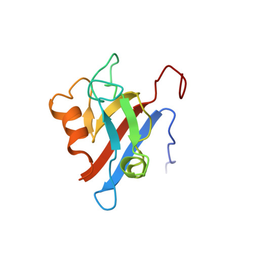
Structure determination and ligand interactions of the PDZ2b domain of PTP-Bas (hPTP1E): Splicing induced modulation of ligand specificity.
Kachel, N., Erdmann, K.S., Kremer, W., Wolff, P., Gronwald, W., Heumann, R., Kalbitzer, H.R.(2003) J Mol Biol 334: 143-155
- PubMed: 14596806
- DOI: https://doi.org/10.1016/j.jmb.2003.09.026
- Primary Citation of Related Structures:
1Q7X - PubMed Abstract:
Two versions of the PDZ2 domain of the protein tyrosine phosphatase PTP-Bas/human PTP-BL are generated by alternative splicing. The domains differ by the insertion of five amino acid residues and their affinity to the tumour suppressor protein APC. Whereas PDZ2a is able to bind APC in the nanomolar range, PDZ2b shows no apparent interaction with APC. Here the solution structure of the splicing variant of PDZ2 with the insertion has been determined using 2D and 3D heteronuclear NMR experiments. The structural reason for the changed binding specificity is the reorientation of the loop with extra five amino acid residues, which folds back onto beta-strands two and three. In addition the side-chain of Lys32 closes the binding site of the APC binding protein and the two helices, especially alpha-helix 2, change their relative position to the protein core. Consecutively, the binding site is sterically no longer fully accessible. From the NMR-titration studies with a C-terminal APC-peptide the affinity of the peptide with the protein can be estimated as 540(+/-40)microM. The binding site encompasses part of the analogous binding site of PDZ2a as already described previously, yet specific interaction sites are abolished by the insertion of amino acids in PDZ2b. As shown by high-affinity chromatography, GST-PDZ2b and GST-PDZ2a bind to phosphatidylinositol 4,5-bisphosphate (PIP(2)) micelles with a dissociation constant K(D) of 21 microM and 55 microM, respectively. In line with these data PDZ2b binds isolated, dissolved PIP(2) and PIP(3) (phosphatidylinositol 3,4,5-trisphosphate) molecules specifically with a lower K(D) of 230(+/-20)microM as detected by NMR spectroscopy. The binding site could be located by our studies and involves the residues Ile24, Val26, Val70, Asn71, Gly77, Ala78, Glu85, Arg88, Gly91 and Gln92. PIP(2) and PIP(3) binding takes place in the groove of the PDZ domain that is normally part of the APC binding site.
Organizational Affiliation:
Institut für Biophysik und Physikalische Biochemie, Universität Regensburg, Universitätsstr. 31, D-93053, Regensburg, Germany.