1Q7L

Zn-binding domain of the T347G mutant of human aminoacylase-I


Experimental Data Snapshot

  • Method: X-RAY DIFFRACTION
  • Resolution: 1.40 Å
  • R-Value Free: 0.172 
  • R-Value Work: 0.133 
  • R-Value Observed: 0.133 

wwPDB Validation   3D Report Full Report


This is version 1.4 of the entry. See complete history


Literature

Essential roles of zinc ligation and enzyme dimerization for catalysis in the aminoacylase-1/M20 family.

Lindner, H.A.Lunin, V.V.Alary, A.Hecker, R.Cygler, M.Menard, R.

(2003) J Biol Chem 278: 44496-44504

  • DOI: https://doi.org/10.1074/jbc.M304233200
  • Primary Citation of Related Structures:  
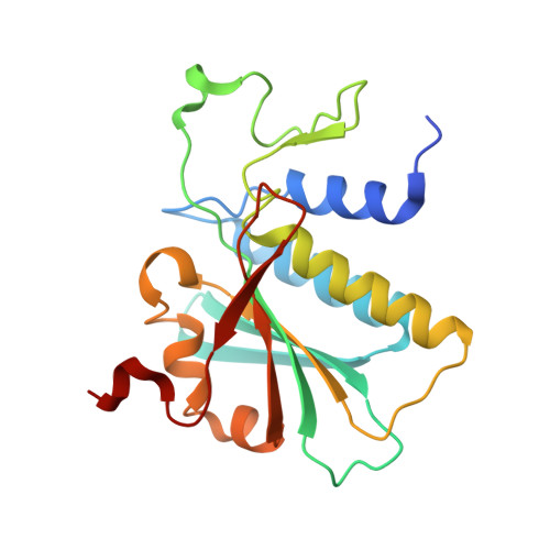
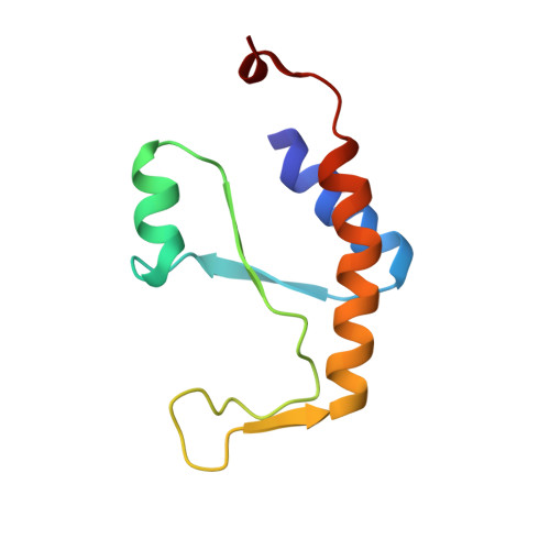
    1Q7L

  • PubMed Abstract: 

    Members of the aminoacylase-1 (Acy1)/M20 family of aminoacylases and exopeptidases exist as either monomers or homodimers. They contain a zinc-binding domain and a second domain mediating dimerization in the latter case. The roles that both domains play in catalysis have been investigated for human Acy1 (hAcy1) by x-ray crystallography and by site-directed mutagenesis. Structure comparison of the dinuclear zinc center in a mutant of hAcy1 reported here with dizinc centers in related enzymes points to a difference in zinc ligation in the Acy1/M20 family. Mutational analysis supports catalytic roles of zinc ions, a vicinal glutamate, and a histidine from the dimerization domain. By complementing different active site mutants of hAcy1, we show that catalysis occurs at the dimer interface. Reinterpretation of the structure of a monomeric homolog, peptidase V, reveals that a domain insertion mimics dimerization. We conclude that monomeric and dimeric Acy1/M20 family members share a unique active site architecture involving both enzyme domains. The study may provide means to improve homologous carboxypeptidase G2 toward application in antibody-directed enzyme prodrug therapy.


  • Organizational Affiliation

    Biotechnology Research Institute, National Research Council of Canada, Montréal, Québec H4P 2R2, Canada.


Macromolecules
Find similar proteins by:  (by identity cutoff)  |  3D Structure
Entity ID: 1
MoleculeChains Sequence LengthOrganismDetailsImage
Aminoacylase-1
A, C
198Homo sapiensMutation(s): 0 
Gene Names: ACY1
EC: 3.5.1.14
UniProt & NIH Common Fund Data Resources
Find proteins for Q03154 (Homo sapiens)
Explore Q03154 
Go to UniProtKB:  Q03154
PHAROS:  Q03154
GTEx:  ENSG00000243989 
Entity Groups  
Sequence Clusters30% Identity50% Identity70% Identity90% Identity95% Identity100% Identity
UniProt GroupQ03154
Sequence Annotations
Expand
  • Reference Sequence
Find similar proteins by:  (by identity cutoff)  |  3D Structure
Entity ID: 2
MoleculeChains Sequence LengthOrganismDetailsImage
Aminoacylase-1
B, D
88Homo sapiensMutation(s): 1 
Gene Names: ACY1
EC: 3.5.1.14
UniProt & NIH Common Fund Data Resources
Find proteins for Q03154 (Homo sapiens)
Explore Q03154 
Go to UniProtKB:  Q03154
PHAROS:  Q03154
GTEx:  ENSG00000243989 
Entity Groups  
Sequence Clusters30% Identity50% Identity70% Identity90% Identity95% Identity100% Identity
UniProt GroupQ03154
Sequence Annotations
Expand
  • Reference Sequence
Experimental Data & Validation

Experimental Data

  • Method: X-RAY DIFFRACTION
  • Resolution: 1.40 Å
  • R-Value Free: 0.172 
  • R-Value Work: 0.133 
  • R-Value Observed: 0.133 
  • Space Group: P 21 21 21
Unit Cell:
Length ( Å )Angle ( ˚ )
a = 53.528α = 90
b = 67.249β = 90
c = 146.479γ = 90
Software Package:
Software NamePurpose
REFMACrefinement
HKL-2000data reduction
SCALEPACKdata scaling
SOLVEphasing

Structure Validation

View Full Validation Report


Entry History 

Deposition Data

Revision History  (Full details and data files)

  • Version 1.0: 2004-01-20
    Type: Initial release
  • Version 1.1: 2008-04-29
    Changes: Version format compliance
  • Version 1.2: 2011-07-13
    Changes: Version format compliance
  • Version 1.3: 2021-10-27
    Changes: Database references, Derived calculations
  • Version 1.4: 2024-02-14
    Changes: Data collection