1Q5R

The Rhodococcus 20S proteasome with unprocessed pro-peptides


Experimental Data Snapshot

  • Method: X-RAY DIFFRACTION
  • Resolution: 3.10 Å
  • R-Value Free: 0.246 
  • R-Value Work: 0.229 
  • R-Value Observed: 0.229 

wwPDB Validation   3D Report Full Report


This is version 1.4 of the entry. See complete history


Literature

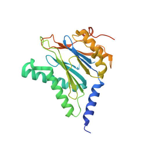
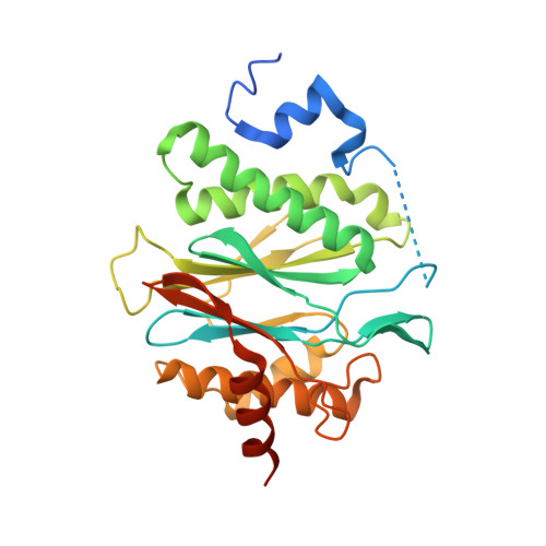
Crystal structures of the Rhodococcus proteasome with and without its pro-peptides: implications for the role of the pro-peptide in proteasome assembly.

Kwon, Y.D.Nagy, I.Adams, P.D.Baumeister, W.Jap, B.K.

(2004) J Mol Biol 335: 233-245

  • DOI: https://doi.org/10.1016/j.jmb.2003.08.029
  • Primary Citation of Related Structures:  
    1Q5Q, 1Q5R

  • PubMed Abstract: 

    To understand the role of the pro-peptide in proteasome assembly, we have determined structures of the Rhodococcus proteasome and a mutant form that prevents the autocatalytic removal of its pro-peptides. The structures reveal that the pro-peptide acts as an assembly-promoting factor by linking its own beta-subunit to two adjacent alpha-subunits, thereby providing a molecular explanation for the observed kinetics of proteasome assembly. The Rhodococcus proteasome has been found to have a substantially smaller contact region between alpha-subunits compared to those regions in the proteasomes of Thermoplasma, yeast, and mammalian cells, suggesting that a smaller contact area between alpha-subunits is likely the structural basis for the Rhodococcus alpha-subunits not assembling into alpha-rings when expressed alone. Analysis of all available beta-subunit structures shows that the contact area between beta-subunits within a beta-ring is not sufficient for beta-ring self-assembly without the additional contact provided by the alpha-ring. This appears to be a fail-safe mechanism ensuring that the active sites on the beta-subunits are activated only after proteasome assembly is complete.


  • Organizational Affiliation

    Graduate Group in Comparative Biochemistry, University of California, Berkeley, CA 94720, USA.


Macromolecules
Find similar proteins by:  (by identity cutoff)  |  3D Structure
Entity ID: 1
MoleculeChains Sequence LengthOrganismDetailsImage
proteasome alpha-type subunit 1
A, B, C, D, E
A, B, C, D, E, F, G
259Rhodococcus erythropolisMutation(s): 0 
Gene Names: PRCA(1)
EC: 3.4.25.1
UniProt
Find proteins for Q53080 (Rhodococcus erythropolis)
Explore Q53080 
Go to UniProtKB:  Q53080
Entity Groups  
Sequence Clusters30% Identity50% Identity70% Identity90% Identity95% Identity100% Identity
UniProt GroupQ53080
Sequence Annotations
Expand
  • Reference Sequence
Find similar proteins by:  (by identity cutoff)  |  3D Structure
Entity ID: 2
MoleculeChains Sequence LengthOrganismDetailsImage
proteasome beta-type subunit 1
H, I, J, K, L
H, I, J, K, L, M, N
294Rhodococcus erythropolisMutation(s): 1 
Gene Names: PRCB(1)
UniProt
Find proteins for Q53079 (Rhodococcus erythropolis)
Explore Q53079 
Go to UniProtKB:  Q53079
Entity Groups  
Sequence Clusters30% Identity50% Identity70% Identity90% Identity95% Identity100% Identity
UniProt GroupQ53079
Sequence Annotations
Expand
  • Reference Sequence
Experimental Data & Validation

Experimental Data

  • Method: X-RAY DIFFRACTION
  • Resolution: 3.10 Å
  • R-Value Free: 0.246 
  • R-Value Work: 0.229 
  • R-Value Observed: 0.229 
  • Space Group: C 2 2 21
Unit Cell:
Length ( Å )Angle ( ˚ )
a = 152.094α = 90
b = 212.365β = 90
c = 250.213γ = 90
Software Package:
Software NamePurpose
CNSrefinement
DENZOdata reduction
SCALEPACKdata scaling
CNSphasing

Structure Validation

View Full Validation Report


Entry History 

Deposition Data

Revision History  (Full details and data files)

  • Version 1.0: 2003-12-16
    Type: Initial release
  • Version 1.1: 2008-04-29
    Changes: Version format compliance
  • Version 1.2: 2011-07-13
    Changes: Version format compliance
  • Version 1.3: 2021-10-27
    Changes: Database references
  • Version 1.4: 2024-02-14
    Changes: Data collection