1Q5L

NMR structure of the substrate binding domain of DnaK bound to the peptide NRLLLTG


Experimental Data Snapshot

  • Method: SOLUTION NMR
  • Conformers Calculated: 60 
  • Conformers Submitted: 15 
  • Selection Criteria: structures with acceptable covalent geometry,structures with the least restraint violations,structures with the lowest energy 

wwPDB Validation   3D Report Full Report


This is version 1.3 of the entry. See complete history


Literature

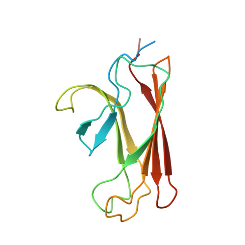
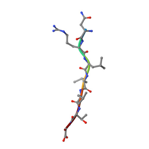
The solution structure of the bacterial HSP70 chaperone protein domain DnaK(393-507) in complex with the peptide NRLLLTG.

Stevens, S.Y.Cai, S.Pellecchia, M.Zuiderweg, E.R.

(2003) Protein Sci 12: 2588-2596

  • DOI: https://doi.org/10.1110/ps.03269103
  • Primary Citation of Related Structures:  
    1Q5L

  • PubMed Abstract: 

    The Hsp70 family of molecular chaperones participates in a number of cellular processes, including binding to nascent polypeptide chains and assistance in protein (re)folding and degradation. We present the solution structure of the substrate binding domain (residues 393-507) of the Escherichia coli Hsp70, DnaK, that is bound to the peptide NRLLLTG and compare it to the crystal structure of DnaK(389-607) bound to the same peptide. The construct discussed here does not contain the alpha-helical domain that characterizes earlier published peptide-bound structures of the Hsp70s. It is established that removing the alpha-helical domain in its entirety does not affect the primary interactions or structure of the DnaK(393-507) in complex with the peptide NRLLLTG. In particular, the arch that protects the substrate-binding cleft is also formed in the absence of the helical lid. 15N-relaxation measurements show that the peptide-bound form of DnaK(393-507) is relatively rigid. As compared to the peptide-free state, the peptide-bound state of the domain shows distinct, widespread, and contiguous differences in structure extending toward areas previously defined as important to the allosteric regulation of the Hsp70 chaperones.


  • Organizational Affiliation

    Biophysics Research Division, University of Michigan, Ann Arbor, Michigan 48109-1055, USA.


Macromolecules
Find similar proteins by:  (by identity cutoff)  |  3D Structure
Entity ID: 1
MoleculeChains Sequence LengthOrganismDetailsImage
Chaperone protein dnaK135Escherichia coliMutation(s): 0 
Gene Names: DNAK
UniProt
Find proteins for P0A6Y8 (Escherichia coli (strain K12))
Explore P0A6Y8 
Go to UniProtKB:  P0A6Y8
Entity Groups  
Sequence Clusters30% Identity50% Identity70% Identity90% Identity95% Identity100% Identity
UniProt GroupP0A6Y8
Sequence Annotations
Expand
  • Reference Sequence

Find similar proteins by:  Sequence   |   3D Structure  

Entity ID: 2
MoleculeChains Sequence LengthOrganismDetailsImage
peptide NRLLLTG7N/AMutation(s): 0 
Sequence Annotations
Expand
  • Reference Sequence
Experimental Data & Validation

Experimental Data

  • Method: SOLUTION NMR
  • Conformers Calculated: 60 
  • Conformers Submitted: 15 
  • Selection Criteria: structures with acceptable covalent geometry,structures with the least restraint violations,structures with the lowest energy 

Structure Validation

View Full Validation Report


Entry History 

Deposition Data

Revision History  (Full details and data files)

  • Version 1.0: 2003-11-04
    Type: Initial release
  • Version 1.1: 2008-04-29
    Changes: Version format compliance
  • Version 1.2: 2011-07-13
    Changes: Version format compliance
  • Version 1.3: 2022-03-02
    Changes: Database references, Derived calculations