1Q57

The Crystal Structure of the Bifunctional Primase-Helicase of Bacteriophage T7


Experimental Data Snapshot

  • Method: X-RAY DIFFRACTION
  • Resolution: 3.45 Å
  • R-Value Free: 0.326 
  • R-Value Work: 0.299 
  • R-Value Observed: 0.301 

wwPDB Validation   3D Report Full Report


This is version 1.4 of the entry. See complete history


Literature

The Crystal Structure of the Bifunctional Primase-Helicase of Bacteriophage T7

Toth, E.A.Li, Y.Sawaya, M.R.Cheng, Y.Ellenberger, T.

(2003) Mol Cell 12: 1113-1123

  • DOI: https://doi.org/10.1016/s1097-2765(03)00442-8
  • Primary Citation of Related Structures:  
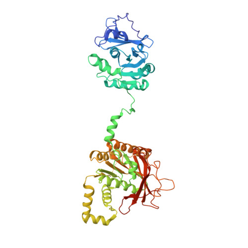
    1Q57

  • PubMed Abstract: 

    Within minutes after infecting Escherichia coli, bacteriophage T7 synthesizes many copies of its genomic DNA. The lynchpin of the T7 replication system is a bifunctional primase-helicase that unwinds duplex DNA at the replication fork while initiating the synthesis of Okazaki fragments on the lagging strand. We have determined a 3.45 A crystal structure of the T7 primase-helicase that shows an articulated arrangement of the primase and helicase sites. The crystallized primase-helicase is a heptamer with a crown-like shape, reflecting an intimate packing of helicase domains into a ring that is topped with loosely arrayed primase domains. This heptameric isoform can accommodate double-stranded DNA in its central channel, which nicely explains its recently described DNA remodeling activity. The double-jointed structure of the primase-helicase permits a free range of motion for the primase and helicase domains that suggests how the continuous unwinding of DNA at the replication fork can be periodically coupled to Okazaki fragment synthesis.


  • Organizational Affiliation

    Department of Biological Chemistry and Molecular Pharmacology, Harvard Medical School, 240 Longwood Avenue, Boston, MA 02115, USA.


Macromolecules
Find similar proteins by:  (by identity cutoff)  |  3D Structure
Entity ID: 1
MoleculeChains Sequence LengthOrganismDetailsImage
DNA primase/helicase
A, B, C, D, E
A, B, C, D, E, F, G
503Escherichia phage T7Mutation(s): 2 
Gene Names: 4
EC: 2.7.7
UniProt
Find proteins for P03692 (Escherichia phage T7)
Explore P03692 
Go to UniProtKB:  P03692
Entity Groups  
Sequence Clusters30% Identity50% Identity70% Identity90% Identity95% Identity100% Identity
UniProt GroupP03692
Sequence Annotations
Expand
  • Reference Sequence
Experimental Data & Validation

Experimental Data

  • Method: X-RAY DIFFRACTION
  • Resolution: 3.45 Å
  • R-Value Free: 0.326 
  • R-Value Work: 0.299 
  • R-Value Observed: 0.301 
  • Space Group: P 1 21 1
Unit Cell:
Length ( Å )Angle ( ˚ )
a = 117.179α = 90
b = 171.567β = 99.86
c = 118.581γ = 90
Software Package:
Software NamePurpose
REFMACrefinement
HKL-2000data reduction
SCALEPACKdata scaling
SHARPphasing

Structure Validation

View Full Validation Report


Entry History 

Deposition Data

Revision History  (Full details and data files)

  • Version 1.0: 2003-11-25
    Type: Initial release
  • Version 1.1: 2008-04-29
    Changes: Version format compliance
  • Version 1.2: 2011-07-13
    Changes: Advisory, Version format compliance
  • Version 1.3: 2021-10-27
    Changes: Database references
  • Version 1.4: 2024-02-14
    Changes: Data collection, Refinement description