1Q4T

crystal structure of 4-hydroxybenzoyl CoA thioesterase from Arthrobacter sp. strain SU complexed with 4-hydroxyphenyl CoA


Experimental Data Snapshot

  • Method: X-RAY DIFFRACTION
  • Resolution: 1.60 Å
  • R-Value Free: 0.219 
  • R-Value Work: 0.179 
  • R-Value Observed: 0.180 

wwPDB Validation   3D Report Full Report


Ligand Structure Quality Assessment 


This is version 1.5 of the entry. See complete history


Literature

The Structure of 4-Hydroxybenzoyl-CoA Thioesterase from Arthrobacter sp. strain SU

Thoden, J.B.Zhuang, Z.Dunaway-Mariano, D.Holden, H.M.

(2003) J Biol Chem 278: 43709-43716

  • DOI: https://doi.org/10.1074/jbc.M308198200
  • Primary Citation of Related Structures:  
    1Q4S, 1Q4T, 1Q4U

  • PubMed Abstract: 

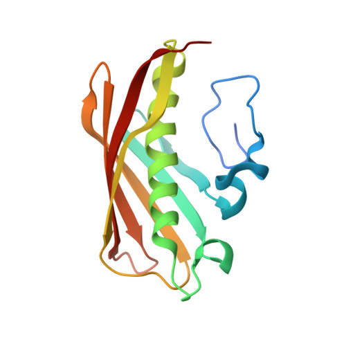
    The 4-chlorobenzoyl-CoA dehalogenation pathway in certain Arthrobacter and Pseudomonas bacterial species contains three enzymes: a ligase, a dehalogenase, and a thioesterase. Here we describe the high resolution x-ray crystallographic structure of the 4-hydroxybenzoyl-CoA thioesterase from Arthrobacter sp. strain SU. The tetrameric enzyme is a dimer of dimers with each subunit adopting the so-called "hot dog fold" composed of six strands of anti-parallel beta-sheet flanked on one side by a rather long alpha-helix. The dimers come together to form the tetramer with their alpha-helices facing outwards. This quaternary structure is in sharp contrast to that previously observed for the 4-hydroxybenzoyl-CoA thioesterase from Pseudomonas species strain CBS-3, whereby the dimers forming the tetramer pack with their alpha-helices projecting toward the interfacial region. In the Arthrobacter thioesterase, each of the four active sites is formed by three of the subunits of the tetramer. On the basis of both structural and kinetic data, it appears that Glu73 is the active site base in the Arthrobacter thioesterase. Remarkably, this residue is located on the opposite side of the substrate-binding pocket compared with that observed for the Pseudomonas enzyme. Although these two bacterial thioesterases demonstrate equivalent catalytic efficiencies, substrate specificities, and metabolic functions, their quaternary structures, CoA-binding sites, and catalytic platforms are decidedly different.


  • Organizational Affiliation

    Department of Biochemistry, University of Wisconsin, Madison, Wisconsin 53706-1544, USA.


Macromolecules
Find similar proteins by:  (by identity cutoff)  |  3D Structure
Entity ID: 1
MoleculeChains Sequence LengthOrganismDetailsImage
Thioesterase
A, B
151Arthrobacter sp. SUMutation(s): 0 
Gene Names: FCBC
EC: 3.1.2.23
UniProt
Find proteins for Q04416 (Arthrobacter sp)
Explore Q04416 
Go to UniProtKB:  Q04416
Entity Groups  
Sequence Clusters30% Identity50% Identity70% Identity90% Identity95% Identity100% Identity
UniProt GroupQ04416
Sequence Annotations
Expand
  • Reference Sequence
Small Molecules
Ligands 3 Unique
IDChains Name / Formula / InChI Key2D Diagram3D Interactions
4CO
Query on 4CO

Download Ideal Coordinates CCD File 
E [auth A],
I [auth B]
4-HYDROXYPHENACYL COENZYME A
C29 H42 N7 O18 P3 S
ZQLMPRRTUJBILA-VXAHOBLNSA-N
EDO
Query on EDO

Download Ideal Coordinates CCD File 
F [auth A],
J [auth B],
K [auth B]
1,2-ETHANEDIOL
C2 H6 O2
LYCAIKOWRPUZTN-UHFFFAOYSA-N
CL
Query on CL

Download Ideal Coordinates CCD File 
C [auth A],
D [auth A],
G [auth B],
H [auth B]
CHLORIDE ION
Cl
VEXZGXHMUGYJMC-UHFFFAOYSA-M
Binding Affinity Annotations 
IDSourceBinding Affinity
4CO Binding MOAD:  1Q4T Ki: 3 (nM) from 1 assay(s)
Experimental Data & Validation

Experimental Data

  • Method: X-RAY DIFFRACTION
  • Resolution: 1.60 Å
  • R-Value Free: 0.219 
  • R-Value Work: 0.179 
  • R-Value Observed: 0.180 
  • Space Group: P 32 2 1
Unit Cell:
Length ( Å )Angle ( ˚ )
a = 112.5α = 90
b = 112.5β = 90
c = 60.6γ = 120
Software Package:
Software NamePurpose
TNTrefinement
SAINTdata reduction
CNSrefinement
FRAMBOdata collection
SAINTdata scaling
CNSphasing

Structure Validation

View Full Validation Report


Ligand Structure Quality Assessment 


Entry History 

Deposition Data

Revision History  (Full details and data files)

  • Version 1.0: 2003-09-23
    Type: Initial release
  • Version 1.1: 2008-04-29
    Changes: Version format compliance
  • Version 1.2: 2011-07-13
    Changes: Derived calculations, Source and taxonomy, Version format compliance
  • Version 1.3: 2017-10-11
    Changes: Refinement description
  • Version 1.4: 2019-07-24
    Changes: Data collection, Refinement description
  • Version 1.5: 2024-02-14
    Changes: Data collection, Database references, Derived calculations