1Q4K

The polo-box domain of Plk1 in complex with a phospho-peptide


Experimental Data Snapshot

  • Method: X-RAY DIFFRACTION
  • Resolution: 2.30 Å
  • R-Value Free: 0.312 
  • R-Value Work: 0.245 
  • R-Value Observed: 0.248 

wwPDB Validation   3D Report Full Report


This is version 1.2 of the entry. See complete history


Literature

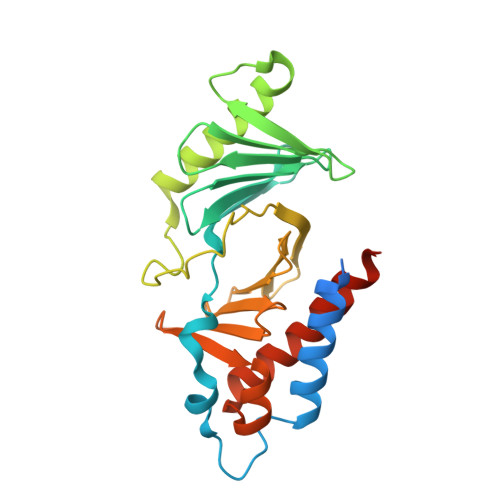
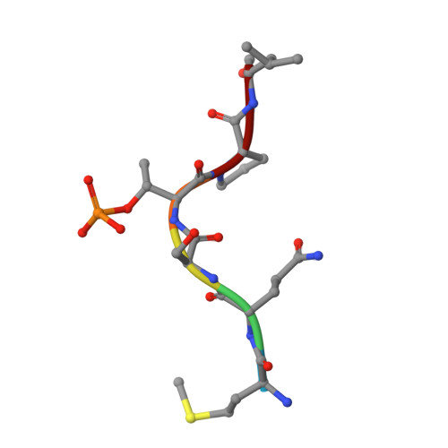
The crystal structure of the human polo-like kinase-1 polo box domain and its phospho-peptide complex.

Cheng, K.Y.Lowe, E.D.Sinclair, J.Nigg, E.A.Johnson, L.N.

(2003) EMBO J 22: 5757-5768

  • DOI: https://doi.org/10.1093/emboj/cdg558
  • Primary Citation of Related Structures:  
    1Q4K, 1Q4O

  • PubMed Abstract: 

    Human polo-like kinase Plk1 localizes to the centrosomes, kinetochores and central spindle structures during mitosis. It plays an essential role in promoting mitosis and cytokinesis through phosphorylation of a number of different substrates. Kinase activity is regulated by a conserved C-terminal domain, termed the polo box domain (PBD), which acts both as an autoinhibitory domain and as a subcellular localization domain. We have determined the crystal structure of Plk1 PBD (residues 367-603) to 2.2 A resolution and the structure of a phospho-peptide-PBD (residues 345-603) complex to 2.3 A resolution. The two polo boxes of the PBD exhibit identical folds based on a six-stranded beta-sheet and an alpha-helix, despite only 12% sequence identity. The phospho-peptide binds at a site between the two polo boxes. It makes a short antiparallel beta-sheet connection and critical contacts to residues Trp414, Leu490, His538 and Lys540. Most of these residues had been shown to be important for biological activity through mutational studies. The results provide an explanation for phospho-peptide recognition and create the basis for new functional studies.


  • Organizational Affiliation

    Laboratory of Molecular Biophysics, Department of Biochemistry, University of Oxford, South Parks Road, Oxford OX1 3QU, UK.


Macromolecules
Find similar proteins by:  (by identity cutoff)  |  3D Structure
Entity ID: 1
MoleculeChains Sequence LengthOrganismDetailsImage
Serine/threonine-protein kinase PLKA [auth B],
B [auth A],
C
259Homo sapiensMutation(s): 0 
Gene Names: PLK1
EC: 2.7.1
UniProt & NIH Common Fund Data Resources
Find proteins for P53350 (Homo sapiens)
Explore P53350 
Go to UniProtKB:  P53350
PHAROS:  P53350
GTEx:  ENSG00000166851 
Entity Groups  
Sequence Clusters30% Identity50% Identity70% Identity90% Identity95% Identity100% Identity
UniProt GroupP53350
Sequence Annotations
Expand
  • Reference Sequence

Find similar proteins by:  Sequence   |   3D Structure  

Entity ID: 2
MoleculeChains Sequence LengthOrganismDetailsImage
Phospho-peptide sequence Met.Gln.Ser.pThr.Pro.Leu
D, E, F
6N/AMutation(s): 0 
Sequence Annotations
Expand
  • Reference Sequence
Small Molecules
Modified Residues  1 Unique
IDChains TypeFormula2D DiagramParent
TPO
Query on TPO
D, E, F
L-PEPTIDE LINKINGC4 H10 N O6 PTHR
Experimental Data & Validation

Experimental Data

  • Method: X-RAY DIFFRACTION
  • Resolution: 2.30 Å
  • R-Value Free: 0.312 
  • R-Value Work: 0.245 
  • R-Value Observed: 0.248 
  • Space Group: P 1
Unit Cell:
Length ( Å )Angle ( ˚ )
a = 57.071α = 91.45
b = 56.888β = 103.21
c = 85.05γ = 118.5
Software Package:
Software NamePurpose
REFMACrefinement
MOSFLMdata reduction
CCP4data scaling
MOLREPphasing

Structure Validation

View Full Validation Report


Entry History 

Deposition Data

Revision History  (Full details and data files)

  • Version 1.0: 2003-11-11
    Type: Initial release
  • Version 1.1: 2008-04-29
    Changes: Version format compliance
  • Version 1.2: 2011-07-13
    Changes: Version format compliance