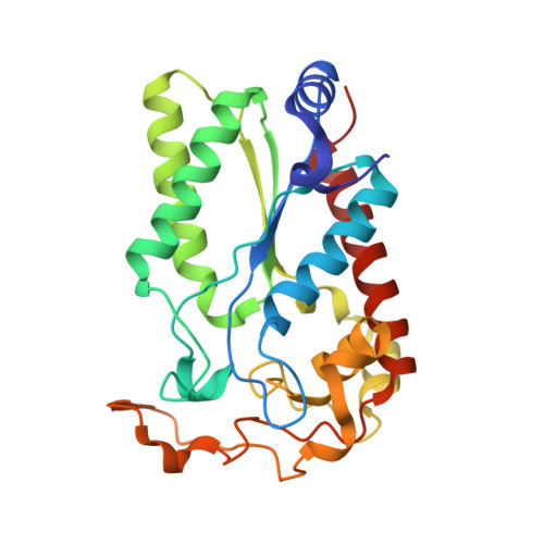
Crystal structure of creatininase from Pseudomonas putida: A novel fold and a case of convergent evolution
Beuth, B., Niefind, K., Schomburg, D.(2003) J Mol Biol 332: 287-301
- PubMed: 12946365
- DOI: https://doi.org/10.1016/s0022-2836(03)00860-x
- Primary Citation of Related Structures:
1Q3K - PubMed Abstract:
Creatinine amidohydrolase (creatininase; EC 3.5.2.10) from Pseudomonas putida, a homohexameric enzyme with a molecular mass of 28.4 kDa per subunit, is a cyclic amidohydrolase catalysing the reversible conversion of creatinine to creatine. The enzyme plays a key role in the bacterial degradation of creatinine. The three-dimensional structure of creatininase from P.putida was determined and refined to 2.1A. The structure shows the six subunits arranged as a trimer of dimers and definitely disproves previous reports that the enzyme has an octameric quaternary structure. Each monomer consists of a central, four-stranded, parallel beta-sheet flanked by two alpha-helices on both sides of the beta-sheet. This topology is unique within the superfamily of amidohydrolases. Moreover, creatininase possesses a novel fold with no close structural relatives within the Protein Data Bank. Each creatininase monomer contains a binuclear zinc centre near the C termini of the beta-strands and the N termini of the main alpha-helices. These zinc ions indicate the location of the active site unambiguously. The active site is entirely buried and is not accessible from the solution without movement of parts of the protein. The two zinc ions are bridged by a water molecule and by an aspartate residue, which acts as a bidentate ligand. They differ from each other in the number and the spatial arrangement of their ligands. One of them is tetrahedrally and the other trigonal-bipyramidally ligated. Using two water molecules of the first coordination sphere as anchor points, a creatinine-water adduct resembling the transition state of the hydrolysation reaction was modelled into the active site. The resulting complex in combination with structural comparisons with other amidohydrolases enabled us to identify the most probable candidate for the catalytic base and to suggest a putative reaction mechanism. Surprisingly these structural comparisons revealed a similarity in the active-site arrangement between creatininase and the hydantoinase-like cyclic amidohydrolases that was unexpected, given the completely unrelated primary and tertiary structures. In particular, the zinc-bridging aspartate residue of creatininase is a spatially and functionally analogue to a carboxylated lysine residue found in dihydroorotase and the hydantoinases. Hence, creatininase and the hydantoinase-like cyclic amidohydrolases represent a further example of convergent evolution within the enzyme class of hydrolases.
Organizational Affiliation:
Universität zu Köln, Institut für Biochemie, Zülpicher Strasse 47, 50674 Köln, Germany.