1Q3A

Crystal structure of the catalytic domain of human matrix metalloproteinase 10


Experimental Data Snapshot

  • Method: X-RAY DIFFRACTION
  • Resolution: 2.10 Å
  • R-Value Free: 0.298 
  • R-Value Work: 0.276 
  • R-Value Observed: 0.278 

wwPDB Validation   3D Report Full Report


Ligand Structure Quality Assessment 


This is version 1.5 of the entry. See complete history


Literature

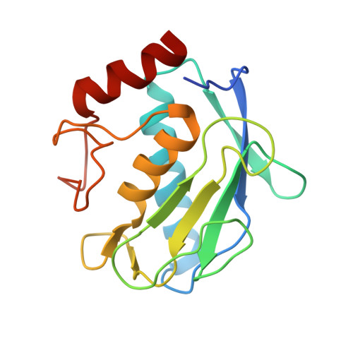
Crystal structure of the catalytic domain of human matrix metalloproteinase 10.

Bertini, I.Calderone, V.Fragai, M.Luchinat, C.Mangani, S.Terni, B.

(2004) J Mol Biol 336: 707-716

  • DOI: https://doi.org/10.1016/j.jmb.2003.12.033
  • Primary Citation of Related Structures:  
    1Q3A

  • PubMed Abstract: 

    The catalytic domain of matrix metalloproteinase-10 (MMP-10) has been expressed in Escherichia coli and its crystal structure solved at 2.1 A resolution. The availability of this structure allowed us to critically examine the small differences existing between the catalytic domains of MMP-3 and MMP-10, which show the highest sequence identity among all MMPs. Furthermore, the binding mode of N-isobutyl-N-[4-methoxyphenylsulfonyl]glycyl hydroxamic acid (NNGH), which is one of the most known commercial inhibitors of MMPs, is described for the first time.


  • Organizational Affiliation

    CERM, University of Florence and FiorGen Foundation, Via Sacconi 6, 50019 Sesto Fiorentino, Florence, Italy. bertini@cerm.unifi.it


Macromolecules
Find similar proteins by:  (by identity cutoff)  |  3D Structure
Entity ID: 1
MoleculeChains Sequence LengthOrganismDetailsImage
Stromelysin-2
A, B, C
165Homo sapiensMutation(s): 1 
Gene Names: MMP10 OR STMY2
EC: 3.4.24.22
UniProt & NIH Common Fund Data Resources
Find proteins for P09238 (Homo sapiens)
Explore P09238 
Go to UniProtKB:  P09238
PHAROS:  P09238
GTEx:  ENSG00000166670 
Entity Groups  
Sequence Clusters30% Identity50% Identity70% Identity90% Identity95% Identity100% Identity
UniProt GroupP09238
Sequence Annotations
Expand
  • Reference Sequence
Small Molecules
Ligands 3 Unique
IDChains Name / Formula / InChI Key2D Diagram3D Interactions
NGH
Query on NGH

Download Ideal Coordinates CCD File 
I [auth A],
O [auth B],
U [auth C]
N-ISOBUTYL-N-[4-METHOXYPHENYLSULFONYL]GLYCYL HYDROXAMIC ACID
C13 H20 N2 O5 S
JIRXORZYIXSWOB-UHFFFAOYSA-N
ZN
Query on ZN

Download Ideal Coordinates CCD File 
D [auth A]
E [auth A]
J [auth B]
K [auth B]
P [auth C]
D [auth A],
E [auth A],
J [auth B],
K [auth B],
P [auth C],
Q [auth C]
ZINC ION
Zn
PTFCDOFLOPIGGS-UHFFFAOYSA-N
CA
Query on CA

Download Ideal Coordinates CCD File 
F [auth A]
G [auth A]
H [auth A]
L [auth B]
M [auth B]
F [auth A],
G [auth A],
H [auth A],
L [auth B],
M [auth B],
N [auth B],
R [auth C],
S [auth C],
T [auth C]
CALCIUM ION
Ca
BHPQYMZQTOCNFJ-UHFFFAOYSA-N
Binding Affinity Annotations 
IDSourceBinding Affinity
NGH BindingDB:  1Q3A Ki: min: 132, max: 133 (nM) from 2 assay(s)
IC50: 6500 (nM) from 1 assay(s)
Experimental Data & Validation

Experimental Data

  • Method: X-RAY DIFFRACTION
  • Resolution: 2.10 Å
  • R-Value Free: 0.298 
  • R-Value Work: 0.276 
  • R-Value Observed: 0.278 
  • Space Group: C 1 2 1
Unit Cell:
Length ( Å )Angle ( ˚ )
a = 117.152α = 90
b = 61.141β = 108.68
c = 68.588γ = 90
Software Package:
Software NamePurpose
REFMACrefinement
SCALAdata scaling
MOLREPphasing
CNSrefinement
CCP4data scaling
MOSFLMdata reduction

Structure Validation

View Full Validation Report


Ligand Structure Quality Assessment 


Entry History 

Deposition Data

Revision History  (Full details and data files)

  • Version 1.0: 2004-04-06
    Type: Initial release
  • Version 1.1: 2008-04-29
    Changes: Version format compliance
  • Version 1.2: 2011-07-13
    Changes: Version format compliance
  • Version 1.3: 2019-07-24
    Changes: Data collection, Refinement description
  • Version 1.4: 2021-10-27
    Changes: Database references, Derived calculations
  • Version 1.5: 2023-08-16
    Changes: Data collection, Refinement description