The Crystal Structure and Mutational Analysis of Human NUDT9
Shen, B.W., Perraud, A.L., Scharenberg, A., Stoddard, B.L.(2003) J Mol Biol 332: 385-398
- PubMed: 12948489
- DOI: https://doi.org/10.1016/s0022-2836(03)00954-9
- Primary Citation of Related Structures:
1Q33, 1QVJ - PubMed Abstract:
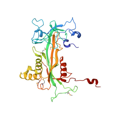
Human ADP-ribose pyrophosphatase NUDT9 belongs to a superfamily of Nudix hydrolases that catabolize potentially toxic compounds in the cell. The enzyme hydrolyzes ADP-ribose (ADPR) to AMP and ribose 5'-phosphate. NUDT9 shares 39% sequence identity with the C-terminal cytoplasmic domain of the ADPR-gated calcium channel TRPM2, which exhibits low but specific enzyme activity. We determined crystal structures of NUDT9 in the presence and in the absence of the reaction product ribose 5'-phosphate. On the basis of these structures and comparison with a bacterial homologue, a model of the substrate complex was built. The structure and activity of a double point mutant (R(229)E(230)F(231) to R(229)I(230)L(231)), which mimics the Nudix signature of the ion channel domain, was determined. Finally, the activities of a pair of additional mutated constructs were compared to the wild-type enzyme. The first corresponds to a minimal Nudix domain missing an N-terminal domain and C-terminal tail; the second disrupts two potential general bases in the active site. NUDT9 contains an N-terminal domain with a novel fold and a catalytic C-terminal Nudix domain. Unlike its closest functional homologue (homodimeric Escherichia coli ADPRase), it is active as a monomer, and the substrate is bound in a cleft between the domains. The structure of the RIL mutant provides structural basis for the reduced activity of the TRPM2 ion channel. The conformation and binding interactions of ADPR substrate are predicted to differ from those observed for E.coli ADPRase; mutation of structurally aligned acidic residues in their active sites produce significantly different effects on catalytic efficiency, indicating that their reaction pathways and mechanisms may have diverged.
Organizational Affiliation:
Division of Basic Sciences, Fred Hutchinson Cancer Research Center, Seattle, WA 98109, USA.