1Q0Z

Crystal structure of aclacinomycin methylesterase (RdmC) with bound product analogue, 10-decarboxymethylaclacinomycin A (DcmA)


Experimental Data Snapshot

  • Method: X-RAY DIFFRACTION
  • Resolution: 1.95 Å
  • R-Value Free: 0.185 
  • R-Value Work: 0.157 
  • R-Value Observed: 0.159 

wwPDB Validation   3D Report Full Report


Ligand Structure Quality Assessment 


This is version 1.4 of the entry. See complete history


Literature

Crystal structure of aclacinomycin methylesterase with bound product analogues: implications for anthracycline recognition and mechanism.

Jansson, A.Niemi, J.Mantsala, P.Schneider, G.

(2003) J Biol Chem 278: 39006-39013

  • DOI: https://doi.org/10.1074/jbc.M304008200
  • Primary Citation of Related Structures:  
    1Q0R, 1Q0Z

  • PubMed Abstract: 

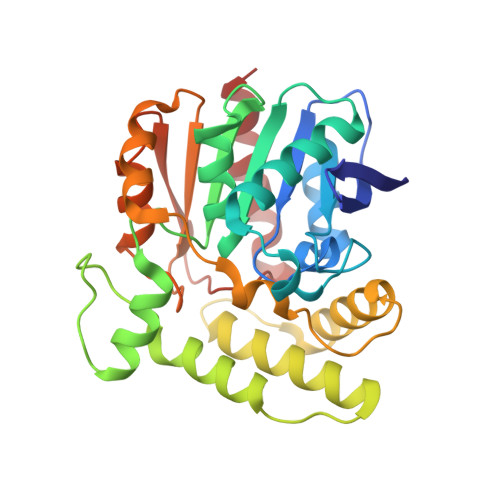
    Aclacinomycin methylesterase (RdmC) is one of the tailoring enzymes that modify the aklavinone skeleton in the biosynthesis of anthracyclines in Streptomyces species. The crystal structures of this enzyme from Streptomyces purpurascens in complex with the product analogues 10-decarboxymethylaclacinomycin T and 10-decarboxymethylaclacinomycin A were determined to nominal resolutions of 1.45 and 1.95 A, respectively. RdmC is built up of two domains. The larger alpha/beta domain shows the common alpha/beta hydrolase fold, whereas the smaller domain is alpha-helical. The active site and substrate binding pocket are located at the interface between the two domains. Decarboxymethylaclacinomycin T and decarboxymethylaclacinomycin A bind close to the catalytic triad (Ser102-His276-Asp248) in a hydrophobic pocket, with the sugar moieties located at the surface of the enzyme. The binding of the ligands is dominated by hydrophobic interactions, and specificity appears to be controlled mainly by the shape of the binding pocket rather than through specific hydrogen bonds. Mechanistic key features consistent with the structure of complexes of RdmC with product analogues are Ser102 acting as nucleophile and transition state stabilization by an oxyanion hole formed by the backbone amides of residues Gly32 and Met103.


  • Organizational Affiliation

    Molecular Structural Biology, Department of Medical Biochemistry and Biophysics, Karolinska Institutet, SE-171 77 Stockholm, Sweden.


Macromolecules
Find similar proteins by:  (by identity cutoff)  |  3D Structure
Entity ID: 1
MoleculeChains Sequence LengthOrganismDetailsImage
aclacinomycin methylesterase298Streptomyces purpurascensMutation(s): 0 
Gene Names: rdmc
UniProt
Find proteins for Q54528 (Streptomyces purpurascens)
Explore Q54528 
Go to UniProtKB:  Q54528
Entity Groups  
Sequence Clusters30% Identity50% Identity70% Identity90% Identity95% Identity100% Identity
UniProt GroupQ54528
Sequence Annotations
Expand
  • Reference Sequence
Small Molecules
Experimental Data & Validation

Experimental Data

  • Method: X-RAY DIFFRACTION
  • Resolution: 1.95 Å
  • R-Value Free: 0.185 
  • R-Value Work: 0.157 
  • R-Value Observed: 0.159 
  • Space Group: P 1 21 1
Unit Cell:
Length ( Å )Angle ( ˚ )
a = 38.106α = 90
b = 84.875β = 99.99
c = 44.389γ = 90
Software Package:
Software NamePurpose
DENZOdata reduction
SCALEPACKdata scaling
SOLVEphasing
ARP/wARPmodel building
REFMACrefinement

Structure Validation

View Full Validation Report


Ligand Structure Quality Assessment 


Entry History 

Deposition Data

Revision History  (Full details and data files)

  • Version 1.0: 2003-11-25
    Type: Initial release
  • Version 1.1: 2008-04-29
    Changes: Version format compliance
  • Version 1.2: 2011-07-13
    Changes: Version format compliance
  • Version 1.3: 2017-10-11
    Changes: Advisory, Refinement description
  • Version 1.4: 2023-08-16
    Changes: Advisory, Data collection, Database references, Derived calculations, Refinement description, Structure summary