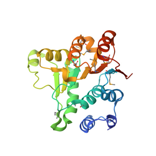
Crystal structure of the BstDEAD N-terminal domain: a novel DEAD protein from Bacillus stearothermophilus
Carmel, A.B., Matthews, B.W.(2004) RNA 10: 66-74
- PubMed: 14681586
- DOI: https://doi.org/10.1261/rna.5134304
- Primary Citation of Related Structures:
1Q0U - PubMed Abstract:
Most cellular processes requiring RNA structure rearrangement necessitate the action of Asp-Glu-Ala-Asp (DEAD) proteins. Members of the family, named originally for the conserved DEAD amino acid sequence, are thought to disrupt RNA structure and facilitate its rearrangement by unwinding short stretches of duplex RNA. BstDEAD is a novel 436 amino acid representative of the DEAD protein family from Bacillus stearothermophilus that contains all eight conserved motifs found in DEAD proteins and is homologous with other members of the family. Here, we describe the 1.85 A resolution structure of the N-terminal domain (residues 1-211) of BstDEAD (BstDEAD-NT). Similar to the corresponding domains of related helicases, BstDEAD-NT adopts a parallel alpha/beta structure with RecA-like topology. In general, the conserved motifs superimpose on closely related DEAD proteins and on more distantly related helicases such as RecA. This affirms the current belief that the core helicase domains, responsible for mechanistic activity, are structurally similar in DEAD proteins. In contrast, however, the so-called Walker A P-loop, which binds the beta- and gamma-phosphates of ATP, adopts a rarely seen "closed" conformation that would sterically block ATP binding. The closed conformation may be indicative of a general regulatory feature among DEAD proteins (and RNA helicases) that differs from that used by DNA helicases. BstDEAD also contains a unique extension of approximately 60 residues at the C terminus that is highly basic, suggesting that it might bind nucleic acids and, in so doing, confer specificity to the helicase activity of the core region.
Organizational Affiliation:
Institute of Molecular Biology, Howard Hughes Medical Institute and Departments of Chemistry and Physics, Eugene, Oregon 97403-1229, USA.