1PZP

TEM-1 Beta-Lactamase in Complex with a Novel, Core-Disrupting, Allosteric Inhibitor


Experimental Data Snapshot

  • Method: X-RAY DIFFRACTION
  • Resolution: 1.45 Å
  • R-Value Free: 0.245 
  • R-Value Work: 0.201 
  • R-Value Observed: 0.203 

wwPDB Validation   3D Report Full Report


Ligand Structure Quality Assessment 


This is version 1.3 of the entry. See complete history


Literature

Allosteric inhibition through core disruption.

Horn, J.R.Shoichet, B.K.

(2004) J Mol Biol 336: 1283-1291

  • DOI: https://doi.org/10.1016/j.jmb.2003.12.068
  • Primary Citation of Related Structures:  
    1PZO, 1PZP

  • PubMed Abstract: 

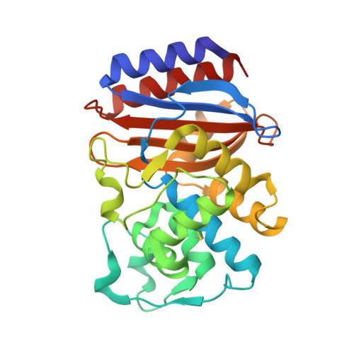
    Although inhibitors typically bind pre-formed sites on proteins, it is theoretically possible to inhibit by disrupting the folded structure of a protein or, in the limit, to bind preferentially to the unfolded state. Equilibria defining how such molecules act are well understood, but structural models for such binding are unknown. Two novel inhibitors of beta-lactamase were found to destabilize the enzyme at high temperatures, but at lower temperatures showed no preference for destabilized mutant enzymes versus stabilized mutants. X-ray crystal structures showed that both inhibitors bound to a cryptic site in beta-lactamase, which the inhibitors themselves created by forcing apart helixes 11 and 12. This opened up a portion of the hydrophobic core of the protein, into which these two inhibitors bind. Although this binding site is 16 A from the center of the active site, the conformational changes were transmitted through a sequence of linked motions to a key catalytic residue, Arg244, which in the complex adopts conformations very different from those in catalytically competent enzyme conformations. These structures offer a detailed view of what has heretofore been a theoretical construct, and suggest the possibility for further design against this novel site.


  • Organizational Affiliation

    Drug Discovery Program, Department of Molecular Pharmacology and Biological Chemistry, Northwestern University School of Medicine, 303 East Chicago Ave, Chicago, IL 60611-3008, USA.


Macromolecules
Find similar proteins by:  (by identity cutoff)  |  3D Structure
Entity ID: 1
MoleculeChains Sequence LengthOrganismDetailsImage
Beta-lactamase TEM263Escherichia coliMutation(s): 1 
Gene Names: bla
EC: 3.5.2.6
UniProt
Find proteins for P62593 (Escherichia coli)
Explore P62593 
Go to UniProtKB:  P62593
Entity Groups  
Sequence Clusters30% Identity50% Identity70% Identity90% Identity95% Identity100% Identity
UniProt GroupP62593
Sequence Annotations
Expand
  • Reference Sequence
Small Molecules
Ligands 1 Unique
IDChains Name / Formula / InChI Key2D Diagram3D Interactions
FTA
Query on FTA

Download Ideal Coordinates CCD File 
B [auth A],
C [auth A]
3-(4-PHENYLAMINO-PHENYLAMINO)-2-(1H-TETRAZOL-5-YL)-ACRYLONITRILE
C16 H13 N7
FLPLCJJGNZGOAW-QXMHVHEDSA-N
Binding Affinity Annotations 
IDSourceBinding Affinity
FTA Binding MOAD:  1PZP Ki: 4.80e+5 (nM) from 1 assay(s)
PDBBind:  1PZP Ki: 4.90e+5 (nM) from 1 assay(s)
Experimental Data & Validation

Experimental Data

  • Method: X-RAY DIFFRACTION
  • Resolution: 1.45 Å
  • R-Value Free: 0.245 
  • R-Value Work: 0.201 
  • R-Value Observed: 0.203 
  • Space Group: P 21 21 21
Unit Cell:
Length ( Å )Angle ( ˚ )
a = 41.553α = 90
b = 60.673β = 90
c = 89.092γ = 90
Software Package:
Software NamePurpose
MOSFLMdata reduction
SCALAdata scaling
AMoREphasing
REFMACrefinement
CCP4data scaling

Structure Validation

View Full Validation Report


Ligand Structure Quality Assessment 


Entry History 

Deposition Data

Revision History  (Full details and data files)

  • Version 1.0: 2004-03-09
    Type: Initial release
  • Version 1.1: 2008-04-29
    Changes: Version format compliance
  • Version 1.2: 2011-07-13
    Changes: Version format compliance
  • Version 1.3: 2021-10-27
    Changes: Database references, Derived calculations