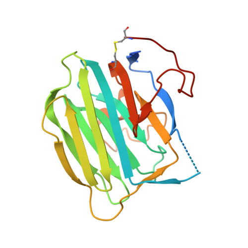
Modulation of agrin function by alternative splicing and Ca2+ binding.
Stetefeld, J., Alexandrescu, A.T., Maciejewski, M.W., Jenny, M., Rathgeb-Szabo, K., Schulthess, T., Landwehr, R., Frank, S., Ruegg, M.A., Kammerer, R.A.(2004) Structure 12: 503-515
- PubMed: 15016366
- DOI: https://doi.org/10.1016/j.str.2004.02.001
- Primary Citation of Related Structures:
1PZ7, 1PZ8, 1PZ9, 1Q56 - PubMed Abstract:
The aggregation of acetylcholine receptors on postsynaptic membranes is a key step in neuromuscular junction development. This process depends on alternatively spliced forms of the proteoglycan agrin with "B-inserts" of 8, 11, or 19 residues in the protein's globular C-terminal domain, G3. Structures of the neural B8 and B11 forms of agrin-G3 were determined by X-ray crystallography. The structure of G3-B0, which lacks inserts, was determined by NMR. The agrin-G3 domain adopts a beta jellyroll fold. The B insert site is flanked by four loops on one edge of the beta sandwich. The loops form a surface that corresponds to a versatile interaction interface in the family of structurally related LNS proteins. NMR and X-ray data indicate that this interaction interface is flexible in agrin-G3 and that flexibility is reduced by Ca(2+) binding. The plasticity of the interaction interface could enable different splice forms of agrin to select between multiple binding partners.
Organizational Affiliation:
Department of Biophysical Chemistry, University of Basel, Klingelbergstrasse 70, CH-4056 Basel, Switzerland. joerg.stetefeld@unibas.ch