


Structure of E. coli Ybab
Kniewel, R., Buglino, J., Chadna, T., Lima, C.D.To be published.
Experimental Data Snapshot
wwPDB Validation 3D Report Full Report

Entity ID: 1 | |||||
|---|---|---|---|---|---|
| Molecule | Chains | Sequence Length | Organism | Details | Image |
| Hypothetical UPF0133 protein ybaB | 109 | Escherichia coli | Mutation(s): 0 Gene Names: YBAB | 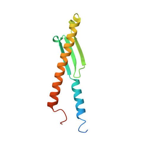 | |
UniProt | |||||
Find proteins for P0A8B5 (Escherichia coli (strain K12)) Explore P0A8B5 Go to UniProtKB: P0A8B5 | |||||
Entity Groups | |||||
| Sequence Clusters | 30% Identity50% Identity70% Identity90% Identity95% Identity100% Identity | ||||
| UniProt Group | P0A8B5 | ||||
Sequence AnnotationsExpand | |||||
| |||||
| Length ( Å ) | Angle ( ˚ ) |
|---|---|
| a = 63.866 | α = 90 |
| b = 84.236 | β = 90 |
| c = 86.636 | γ = 90 |
| Software Name | Purpose |
|---|---|
| DENZO | data reduction |
| SCALEPACK | data scaling |
| SOLVE | phasing |
| RESOLVE | model building |
| REFMAC | refinement |
| RESOLVE | phasing |
RCSB PDB (citation) is hosted by
RCSB PDB is a member of the

