1PTW

The Crystal Structure of AMP-Bound PDE4 Suggests a Mechanism for Phosphodiesterase Catalysis


Experimental Data Snapshot

  • Method: X-RAY DIFFRACTION
  • Resolution: 2.30 Å
  • R-Value Free: 0.274 
  • R-Value Work: 0.229 

wwPDB Validation   3D Report Full Report


Ligand Structure Quality Assessment 


This is version 1.3 of the entry. See complete history


Literature

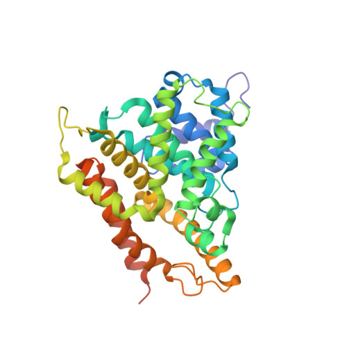
The crystal structure of AMP-bound PDE4 suggests a mechanism for phosphodiesterase catalysis

Huai, Q.Colicelli, J.Ke, H.

(2003) Biochemistry 42: 13220-13226

  • DOI: https://doi.org/10.1021/bi034653e
  • Primary Citation of Related Structures:  
    1PTW

  • PubMed Abstract: 

    Cyclic nucleotide phosphodiesterases (PDEs) regulate the intracellular concentrations of cyclic 3',5'-adenosine and guanosine monophosphates (cAMP and cGMP, respectively) by hydrolyzing them to AMP and GMP, respectively. Family-selective inhibitors of PDEs have been studied for treatment of various human diseases. However, the catalytic mechanism of cyclic nucleotide hydrolysis by PDEs has remained unclear. We determined the crystal structure of the human PDE4D2 catalytic domain in complex with AMP at 2.4 A resolution. In this structure, two divalent metal ions simultaneously interact with the phosphate group of AMP, implying a binuclear catalysis. In addition, the structure suggested that a hydroxide ion or a water bridging two metal ions may serve as the nucleophile for the hydrolysis of the cAMP phosphodiester bond.


  • Organizational Affiliation

    Department of Biochemistry and Biophysics and Lineberger Comprehensive Cancer Center, The University of North Carolina, Chapel Hill, North Carolina 27599-7260, USA.


Macromolecules
Find similar proteins by:  (by identity cutoff)  |  3D Structure
Entity ID: 1
MoleculeChains Sequence LengthOrganismDetailsImage
cAMP-specific phosphodiesterase PDE4D2
A, B, C, D
360Homo sapiensMutation(s): 0 
Gene Names: PDE4D2
EC: 3.1.4.17
UniProt & NIH Common Fund Data Resources
Find proteins for Q08499 (Homo sapiens)
Explore Q08499 
Go to UniProtKB:  Q08499
PHAROS:  Q08499
GTEx:  ENSG00000113448 
Entity Groups  
Sequence Clusters30% Identity50% Identity70% Identity90% Identity95% Identity100% Identity
UniProt GroupQ08499
Sequence Annotations
Expand
  • Reference Sequence
Small Molecules
Ligands 2 Unique
IDChains Name / Formula / InChI Key2D Diagram3D Interactions
AMP
Query on AMP

Download Ideal Coordinates CCD File 
G [auth A],
J [auth B],
M [auth C],
P [auth D]
ADENOSINE MONOPHOSPHATE
C10 H14 N5 O7 P
UDMBCSSLTHHNCD-KQYNXXCUSA-N
ZN
Query on ZN

Download Ideal Coordinates CCD File 
E [auth A]
F [auth A]
H [auth B]
I [auth B]
K [auth C]
E [auth A],
F [auth A],
H [auth B],
I [auth B],
K [auth C],
L [auth C],
N [auth D],
O [auth D]
ZINC ION
Zn
PTFCDOFLOPIGGS-UHFFFAOYSA-N
Experimental Data & Validation

Experimental Data

  • Method: X-RAY DIFFRACTION
  • Resolution: 2.30 Å
  • R-Value Free: 0.274 
  • R-Value Work: 0.229 
  • Space Group: P 21 21 21
Unit Cell:
Length ( Å )Angle ( ˚ )
a = 99.207α = 90
b = 111.229β = 90
c = 159.65γ = 90
Software Package:
Software NamePurpose
DENZOdata reduction
SCALEPACKdata scaling
AMoREphasing
CNSrefinement

Structure Validation

View Full Validation Report


Ligand Structure Quality Assessment 


Entry History 

Deposition Data

Revision History  (Full details and data files)

  • Version 1.0: 2003-11-11
    Type: Initial release
  • Version 1.1: 2008-04-29
    Changes: Version format compliance
  • Version 1.2: 2011-07-13
    Changes: Version format compliance
  • Version 1.3: 2024-02-14
    Changes: Data collection, Database references, Derived calculations