1PTH

The Structural Basis of Aspirin Activity Inferred from the Crystal Structure of Inactivated Prostaglandin H2 Synthase


Experimental Data Snapshot

  • Method: X-RAY DIFFRACTION
  • Resolution: 3.40 Å
  • R-Value Free: 0.227 
  • R-Value Work: 0.186 
  • R-Value Observed: 0.186 

wwPDB Validation   3D Report Full Report


This is version 2.0 of the entry. See complete history


Literature

The structural basis of aspirin activity inferred from the crystal structure of inactivated prostaglandin H2 synthase.

Loll, P.J.Picot, D.Garavito, R.M.

(1995) Nat Struct Biol 2: 637-643

  • DOI: https://doi.org/10.1038/nsb0895-637
  • Primary Citation of Related Structures:  
    1PTH

  • PubMed Abstract: 

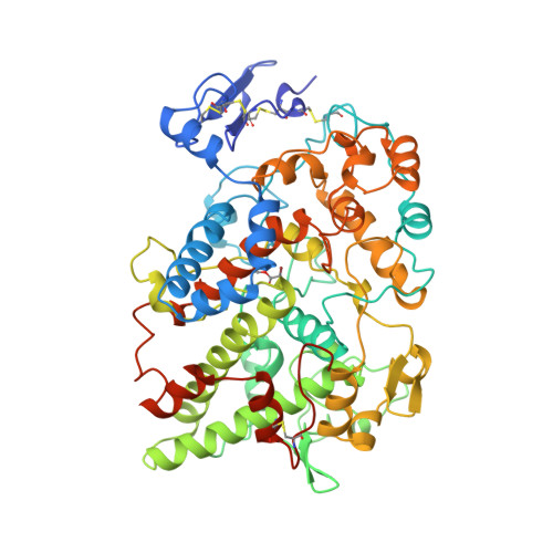
    Aspirin exerts its anti-inflammatory effects through selective acetylation of serine 530 on prostaglandin H2 synthase (PGHS). Here we present the 3.4 A resolution X-ray crystal structure of PGHS isoform-1 inactivated by the potent aspirin analogue 2-bromoacetoxy-benzoic acid. Acetylation by this analogue abolishes cyclooxygenase activity by steric blockage of the active-site channel and not through a large conformational change. We observe two rotameric states of the acetyl-serine side chain which block the channel to different extents, a result which may explain the dissimilar effects of aspirin on the two PGHS isoforms. We also observe the product salicylic acid binding at a site consistent with its antagonistic effect on aspirin activity.


  • Organizational Affiliation

    Department of Biochemistry and Molecular Biology, University of Chicago, Illinois 60637, USA.


Macromolecules
Find similar proteins by:  (by identity cutoff)  |  3D Structure
Entity ID: 1
MoleculeChains Sequence LengthOrganismDetailsImage
PROSTAGLANDIN H2 SYNTHASE-1
A, B
576Ovis ariesMutation(s): 0 
EC: 1.14.99.1
Membrane Entity: Yes 
UniProt
Find proteins for P05979 (Ovis aries)
Explore P05979 
Go to UniProtKB:  P05979
Entity Groups  
Sequence Clusters30% Identity50% Identity70% Identity90% Identity95% Identity100% Identity
UniProt GroupP05979
Sequence Annotations
Expand
  • Reference Sequence
Oligosaccharides

Help

Entity ID: 2
MoleculeChains Length2D Diagram Glycosylation3D Interactions
2-acetamido-2-deoxy-beta-D-glucopyranose-(1-4)-2-acetamido-2-deoxy-beta-D-glucopyranose
C, D
2N-Glycosylation
Glycosylation Resources
GlyTouCan:  G42666HT
GlyCosmos:  G42666HT
GlyGen:  G42666HT
Small Molecules
Ligands 4 Unique
IDChains Name / Formula / InChI Key2D Diagram3D Interactions
HEM
Query on HEM

Download Ideal Coordinates CCD File 
H [auth A],
M [auth B]
PROTOPORPHYRIN IX CONTAINING FE
C34 H32 Fe N4 O4
KABFMIBPWCXCRK-RGGAHWMASA-L
BOG
Query on BOG

Download Ideal Coordinates CCD File 
G [auth A],
L [auth B]
octyl beta-D-glucopyranoside
C14 H28 O6
HEGSGKPQLMEBJL-RKQHYHRCSA-N
NAG
Query on NAG

Download Ideal Coordinates CCD File 
E [auth A],
F [auth A],
J [auth B],
K [auth B]
2-acetamido-2-deoxy-beta-D-glucopyranose
C8 H15 N O6
OVRNDRQMDRJTHS-FMDGEEDCSA-N
SAL
Query on SAL

Download Ideal Coordinates CCD File 
I [auth A],
N [auth B]
2-HYDROXYBENZOIC ACID
C7 H6 O3
YGSDEFSMJLZEOE-UHFFFAOYSA-N
Modified Residues  1 Unique
IDChains TypeFormula2D DiagramParent
0AH
Query on 0AH
A, B
L-PEPTIDE LINKINGC5 H8 Br N O4SER
Binding Affinity Annotations 
IDSourceBinding Affinity
SAL BindingDB:  1PTH Ki: 1.00e+4 (nM) from 1 assay(s)
IC50: min: 4.00e+4, max: 5.00e+5 (nM) from 2 assay(s)
Experimental Data & Validation

Experimental Data

  • Method: X-RAY DIFFRACTION
  • Resolution: 3.40 Å
  • R-Value Free: 0.227 
  • R-Value Work: 0.186 
  • R-Value Observed: 0.186 
  • Space Group: I 2 2 2
Unit Cell:
Length ( Å )Angle ( ˚ )
a = 99.57α = 90
b = 209.8β = 90
c = 235γ = 90
Software Package:
Software NamePurpose
MADNESdata collection
X-PLORmodel building
X-PLORrefinement
MADNESdata reduction
X-PLORphasing

Structure Validation

View Full Validation Report


Entry History 

Deposition Data

Revision History  (Full details and data files)

  • Version 1.0: 1996-04-11
    Type: Initial release
  • Version 1.1: 2008-03-03
    Changes: Version format compliance
  • Version 1.2: 2011-07-13
    Changes: Non-polymer description, Version format compliance
  • Version 2.0: 2020-07-29
    Type: Remediation
    Reason: Carbohydrate remediation
    Changes: Atomic model, Data collection, Derived calculations, Other, Structure summary