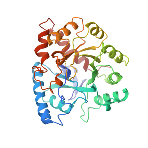
Three-dimensional structure of phosphotriesterase: an enzyme capable of detoxifying organophosphate nerve agents.
Benning, M.M., Kuo, J.M., Raushel, F.M., Holden, H.M.(1994) Biochemistry 33: 15001-15007
- PubMed: 7999757
- DOI: https://doi.org/10.1021/bi00254a008
- Primary Citation of Related Structures:
1PTA - PubMed Abstract:
Organophosphates, such as parathion and paraoxon, constitute the largest class of insecticides currently used in industrialized nations. In addition, many of these compounds are known to inhibit mammalian acetylcholinesterases thereby acting as nerve agents. Consequently, organophosphate-degrading enzymes are of considerable interest in light of their ability to detoxify such compounds. Here we report the three-dimensional structure of such an enzyme, namely, phosphotriesterase, as determined by single crystal X-ray diffraction analysis to 2.1-A resolution. Crystals employed in this investigation belonged to the space group P2(1)2(1)2 with unit cell dimensions of a = 80.3 A, b = 93.4 A, and c = 44.8 A and one molecule per asymmetric unit. The structure was solved by multiple isomorphous replacement with two heavy-atom derivatives and refined to a crystallographic R factor of 18.0%. As observed in various other enzymes, the overall fold of the molecule consists of an alpha/beta barrel with eight strands of parallel beta-pleated sheet. In addition, there are two antiparallel beta-strands at the N-terminus. The molecular model of phosphotriesterase presented here provides the initial structural framework necessary toward understanding the enzyme's broad substrate specificities and its catalytic mechanism.
Organizational Affiliation:
Department of Biochemistry, University of Wisconsin, Madison 53705.