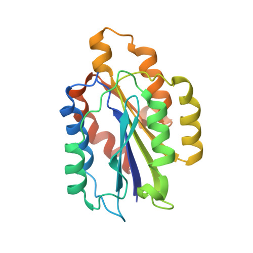
Jararhagin-derived RKKH peptides induce structural changes in alpha1I domain of human integrin alpha1beta1.
Nymalm, Y., Puranen, J.S., Nyholm, T.K.M., Kapyla, J., Kidron, H., Airenne, T.T., Heino, J., Slotte, J.P., Johnson, M.S., Salminen, T.A.(2004) J Biol Chem 279: 7962-7970
- PubMed: 14660600
- DOI: https://doi.org/10.1074/jbc.M312912200
- Primary Citation of Related Structures:
1PT6, 1QCY - PubMed Abstract:
Integrin alpha(1)beta(1) is one of four collagen-binding integrins in humans. Collagens bind to the alphaI domain and in the case of alpha(2)I collagen binding is competitively inhibited by peptides containing the RKKH sequence and derived from the metalloproteinase jararhagin of snake venom from Bothrops jararaca. In alpha(2)I, these peptides bind near the metal ion-dependent adhesion site (MIDAS), where a collagen (I)-like peptide is known to bind; magnesium is required for binding. Published structures of the ligand-bound "open" conformation of alpha(2)I differs significantly from the "closed" conformation seen in the structure of apo-alpha(2)I near MIDAS. Here we show that two peptides, CTRKKHDC and CARKKHDC, derived from jararhagin also bind to alpha(1)I and competitively inhibit collagen I binding. Furthermore, calorimetric and fluorimetric measurements show that the structure of the complex of alpha(1)I with Mg(2+) and CTRKKHDC differs from structure in the absence of peptide. A comparison of the x-ray structure of apo-alpha(1)I ("closed" conformation) and a model structure of the alpha(1)I ("open" conformation) based on the closely related structure of alpha(2)I reveals that the binding site is partially blocked to ligands by Glu(255) and Tyr(285) in the "closed" structure, whereas in the "open" structure helix C is unwound and these residues are shifted, and the "RKKH" peptides fit well when docked. The "open" conformation of alpha(2)I resulting from binding a collagen (I)-like peptide leads to exposure of hydrophobic surface, also seen in the model of alpha(1)I and shown experimentally for alpha(1)I using a fluorescent hydrophobic probe.
Organizational Affiliation:
Department of Biochemistry and Pharmacy, Abo Akademi University, P.O. Box 66, FIN-20521, Turku, Finland.