1PSD

THE ALLOSTERIC LIGAND SITE IN THE VMAX-TYPE COOPERATIVE ENZYME PHOSPHOGLYCERATE DEHYDROGENASE


Experimental Data Snapshot

  • Method: X-RAY DIFFRACTION
  • Resolution: 2.75 Å
  • R-Value Work: 0.177 
  • R-Value Observed: 0.177 

wwPDB Validation   3D Report Full Report


This is version 1.4 of the entry. See complete history


Literature

The allosteric ligand site in the Vmax-type cooperative enzyme phosphoglycerate dehydrogenase.

Schuller, D.J.Grant, G.A.Banaszak, L.J.

(1995) Nat Struct Biol 2: 69-76

  • DOI: https://doi.org/10.1038/nsb0195-69
  • Primary Citation of Related Structures:  
    1PSD

  • PubMed Abstract: 

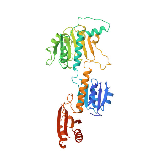
    The crystal structure of the phosphoglycerate dehydrogenase from Escherichia coli is unique among dehydrogenases. It consists of three clearly separate domains connected by flexible hinges. The tetramer has approximate 222 symmetry with the principal contacts between the subunits forming between either the nucleotide binding domains or the regulatory domains. Two slightly different subunit conformations are present which vary only in the orientations of the domains. There is a hinge-like arrangement near the active site cleft and the serine effector site is provided by the regulatory domain of each of two subunits. Interdomain flexibility may play a key role in both catalysis and allosteric inhibition.


  • Organizational Affiliation

    Department of Molecular Biology, University of California, Irvine 92717.


Macromolecules
Find similar proteins by:  (by identity cutoff)  |  3D Structure
Entity ID: 1
MoleculeChains Sequence LengthOrganismDetailsImage
D-3-PHOSPHOGLYCERATE DEHYDROGENASE (PHOSPHOGLYCERATE DEHYDROGENASE)
A, B
409Escherichia coli K-12Mutation(s): 0 
EC: 1.1.1.95
UniProt
Find proteins for P0A9T0 (Escherichia coli (strain K12))
Explore P0A9T0 
Go to UniProtKB:  P0A9T0
Entity Groups  
Sequence Clusters30% Identity50% Identity70% Identity90% Identity95% Identity100% Identity
UniProt GroupP0A9T0
Sequence Annotations
Expand
  • Reference Sequence
Small Molecules
Experimental Data & Validation

Experimental Data

  • Method: X-RAY DIFFRACTION
  • Resolution: 2.75 Å
  • R-Value Work: 0.177 
  • R-Value Observed: 0.177 
  • Space Group: P 21 21 2
Unit Cell:
Length ( Å )Angle ( ˚ )
a = 136.1α = 90
b = 146.3β = 90
c = 53.3γ = 90
Software Package:
Software NamePurpose
X-PLORmodel building
X-PLORrefinement
X-PLORphasing

Structure Validation

View Full Validation Report


Entry History 

Deposition Data

Revision History  (Full details and data files)

  • Version 1.0: 1995-07-31
    Type: Initial release
  • Version 1.1: 2008-03-03
    Changes: Version format compliance
  • Version 1.2: 2011-07-13
    Changes: Derived calculations, Source and taxonomy, Version format compliance
  • Version 1.3: 2017-11-29
    Changes: Derived calculations, Other
  • Version 1.4: 2024-02-14
    Changes: Data collection, Database references, Derived calculations