Three-dimensional structure of the binuclear metal center of phosphotriesterase.
Benning, M.M., Kuo, J.M., Raushel, F.M., Holden, H.M.(1995) Biochemistry 34: 7973-7978
- PubMed: 7794910
- DOI: https://doi.org/10.1021/bi00025a002
- Primary Citation of Related Structures:
1PSC - PubMed Abstract:
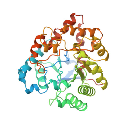
Phosphotriesterase, as isolated from Pseudomonas diminuta, is capable of detoxifying widely used pesticides such as paraoxon and parathion and various mammalian acetylcholinesterase inhibitors. The enzyme requires a binuclear metal center for activity. Recently, the three-dimensional structure of the apoenzyme was solved (Benning et al., 1994) and shown to consist of an alpha/beta-barrel. Here we describe the three-dimensional structure of the holoenzyme, reconstituted with cadmium, as determined by X-ray crystallographic analysis to 2.0-A resolution. Crystals employed in the investigation belonged to the space group C2 with unit cell dimensions of a = 129.5 A, b = 91.4 A, c = 69.4 A, beta = 91.9 degrees, and two subunits in the asymmetric unit. There are significant differences in the three-dimensional architecture of the apo and holo forms of the enzyme such that their alpha-carbon positions superimpose with a root-mean-square deviation of 3.4 A. The binuclear metal center is located at the C-terminus of the beta-barrel with the cadmiums separated by 3.8 A. There are two bridging ligands to the metals: a water molecule (or possibly a hydroxide ion) and a carbamylated lysine residue (Lys 169). The more buried cadmium is surrounded by His 55, His 57, Lys 169, Asp 301, and the bridging water in a trigonal bipyramidal arrangement. The second metal is coordinated in a distorted octahedral geometry by His 201, His 230, Lys 169, the bridging water molecule, and two additional solvents.
Organizational Affiliation:
Institute for Enzyme Research, Graduate School, University of Wisconsin, Madison 53705, USA.