1PSB

Solution structure of calcium loaded S100B complexed to a peptide from N-Terminal regulatory domain of NDR kinase.


Experimental Data Snapshot

  • Method: SOLUTION NMR
  • Conformers Calculated: 128 
  • Conformers Submitted: 20 
  • Selection Criteria: structures with the least restraint violations,structures with the lowest energy 

wwPDB Validation   3D Report Full Report


This is version 1.3 of the entry. See complete history


Literature

Structure of the Ca(2+)/S100B/NDR Kinase Peptide Complex: Insights into S100 Target Specificity and Activation of the Kinase.

Bhattacharya, S.Large, E.Heizmann, C.W.Hemmings, B.Chazin, W.J.

(2003) Biochemistry 42: 14416-14426

  • DOI: https://doi.org/10.1021/bi035089a
  • Primary Citation of Related Structures:  
    1PSB

  • PubMed Abstract: 

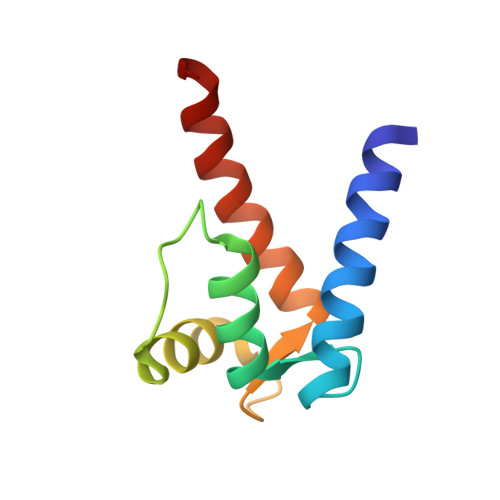
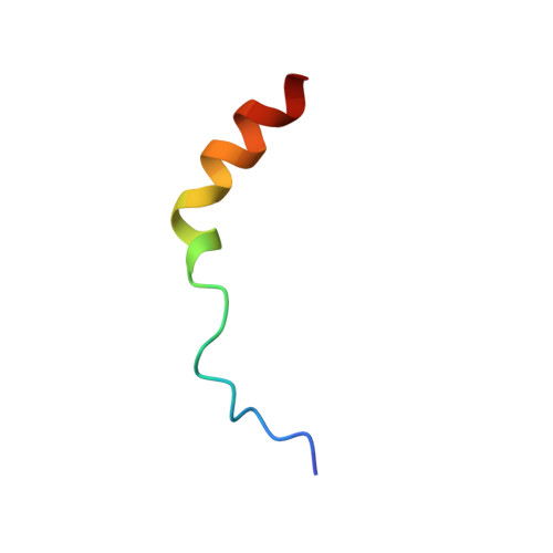
    NDR, a nuclear serine/threonine kinase, belongs to the subfamily of Dbf2 kinases that is critical to the morphology and proliferation of cells. The activity of NDR kinase is modulated in a Ca(2+)/S100B-dependent manner by phosphorylation of Ser281 in the catalytic domain and Thr444 in the C-terminal regulatory domain. S100B, which is a member of the S100 subfamily of EF-hand proteins, binds to a basic/hydrophobic sequence at the junction of the N-terminal regulatory and catalytic domains (NDR(62-87)). Unlike calmodulin-dependent kinases, regulation of NDR by S100B is not associated with direct autoinhibition of the active site, but rather involves a conformational change in the catalytic domain triggered by Ca(2+)/S100B binding to the junction region. To gain further insight into the mechanism of activation of the kinase, studies have been carried out on Ca(2+)/S100B in complex with the intact N-terminal regulatory domain, NDR(1-87). Multidimensional heteronuclear NMR analysis showed that the binding mode and stoichiometry of a peptide fragment of NDR (NDR(62-87)) is the same as for the intact N-terminal regulatory domain. The solution structure of Ca(2+)/S100B and NDR(62-87) has been determined. One target molecule is found to associate with each subunit of the S100B dimer. The peptide adopts three turns of helix in the bound state, and the complex is stabilized by both hydrophobic and electrostatic interactions. These structural studies, in combination with available biochemical data, have been used to develop a model for calcium-induced activation of NDR kinase by S100B.


  • Organizational Affiliation

    Department of Biochemistry, Center for Structural Biology, Vanderbilt University, Nashville, Tennessee 37232-8725, USA.


Macromolecules
Find similar proteins by:  (by identity cutoff)  |  3D Structure
Entity ID: 1
MoleculeChains Sequence LengthOrganismDetailsImage
S-100 protein, beta chain
A, B
91Bos taurusMutation(s): 0 
Gene Names: S100B
UniProt
Find proteins for P02638 (Bos taurus)
Explore P02638 
Go to UniProtKB:  P02638
Entity Groups  
Sequence Clusters30% Identity50% Identity70% Identity90% Identity95% Identity100% Identity
UniProt GroupP02638
Sequence Annotations
Expand
  • Reference Sequence
Find similar proteins by:  (by identity cutoff)  |  3D Structure
Entity ID: 2
MoleculeChains Sequence LengthOrganismDetailsImage
Ndr Ser/Thr kinase-like protein
C, D
26N/AMutation(s): 0 
UniProt & NIH Common Fund Data Resources
Find proteins for Q15208 (Homo sapiens)
Explore Q15208 
Go to UniProtKB:  Q15208
PHAROS:  Q15208
GTEx:  ENSG00000112079 
Entity Groups  
Sequence Clusters30% Identity50% Identity70% Identity90% Identity95% Identity100% Identity
UniProt GroupQ15208
Sequence Annotations
Expand
  • Reference Sequence
Experimental Data & Validation

Experimental Data

  • Method: SOLUTION NMR
  • Conformers Calculated: 128 
  • Conformers Submitted: 20 
  • Selection Criteria: structures with the least restraint violations,structures with the lowest energy 

Structure Validation

View Full Validation Report


Entry History 

Deposition Data

Revision History  (Full details and data files)

  • Version 1.0: 2003-12-16
    Type: Initial release
  • Version 1.1: 2008-04-29
    Changes: Version format compliance
  • Version 1.2: 2011-07-13
    Changes: Version format compliance
  • Version 1.3: 2022-03-02
    Changes: Data collection, Database references, Derived calculations