1POP

X-RAY CRYSTALLOGRAPHIC STRUCTURE OF A PAPAIN-LEUPEPTIN COMPLEX


Experimental Data Snapshot

  • Method: X-RAY DIFFRACTION
  • Resolution: 2.10 Å

wwPDB Validation   3D Report Full Report


This is version 1.5 of the entry. See complete history


Literature

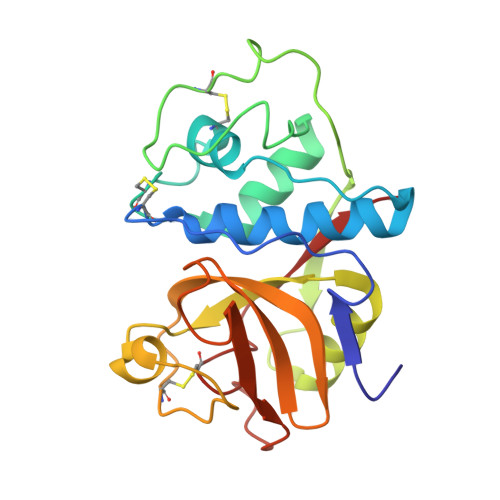
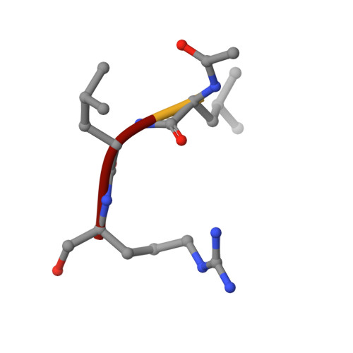
X-ray crystallographic structure of a papain-leupeptin complex.

Schroder, E.Phillips, C.Garman, E.Harlos, K.Crawford, C.

(1993) FEBS Lett 315: 38-42

  • DOI: https://doi.org/10.1016/0014-5793(93)81128-m
  • Primary Citation of Related Structures:  
    1POP

  • PubMed Abstract: 

    The three-dimensional structure of the papain-leupeptin complex has been determined by X-ray crystallography to a resolution of 2.1 A (overall R-factor = 19.8%). The structure indicates that: (i) leupeptin contacts the S subsites of the papain active site and not the S' subsites; (ii) the 'carbonyl' carbon atom of the inhibitor is covalently bound by the Cys-25 sulphur atom of papain and is tetrahedrally coordinated; (iii) the 'carbonyl' oxygen atom of the inhibitor faces the oxyanion hole and makes hydrogen bond contacts with Gln-19 and Cys-25.


  • Organizational Affiliation

    University of Oxford, Laboratory of Molecular Biophysics, UK.


Macromolecules
Find similar proteins by:  (by identity cutoff)  |  3D Structure
Entity ID: 1
MoleculeChains Sequence LengthOrganismDetailsImage
PAPAIN212Carica papayaMutation(s): 0 
EC: 3.4.22.2
UniProt
Find proteins for P00784 (Carica papaya)
Explore P00784 
Go to UniProtKB:  P00784
Entity Groups  
Sequence Clusters30% Identity50% Identity70% Identity90% Identity95% Identity100% Identity
UniProt GroupP00784
Sequence Annotations
Expand
  • Reference Sequence

Find similar proteins by:  Sequence   |   3D Structure  

Entity ID: 2
MoleculeChains Sequence LengthOrganismDetailsImage
LEUPEPTIN4Streptomyces roseusMutation(s): 0 
Sequence Annotations
Expand
  • Reference Sequence
Small Molecules
Ligands 1 Unique
IDChains Name / Formula / InChI Key2D Diagram3D Interactions
MOH
Query on MOH

Download Ideal Coordinates CCD File 
C [auth A]
D [auth A]
E [auth A]
F [auth A]
G [auth A]
C [auth A],
D [auth A],
E [auth A],
F [auth A],
G [auth A],
H [auth A],
I [auth A],
J [auth A],
K [auth A],
L [auth A],
M [auth A],
N [auth A],
O [auth A],
P [auth A],
Q [auth A],
R [auth A],
S [auth A],
T [auth A],
U [auth A],
V [auth A],
W [auth A],
X [auth A],
Y [auth A],
Z [auth B]
METHANOL
C H4 O
OKKJLVBELUTLKV-UHFFFAOYSA-N
Modified Residues  1 Unique
IDChains TypeFormula2D DiagramParent
AR7
Query on AR7
B
PEPTIDE-LIKEC6 H17 N4 O2ARG
Biologically Interesting Molecules (External Reference) 1 Unique
Entity ID: 2
IDChains NameType/Class2D Diagram3D Interactions
PRD_000216
Query on PRD_000216
B
LEUPEPTINOligopeptide / Enzyme inhibitor
Experimental Data & Validation

Experimental Data

  • Method: X-RAY DIFFRACTION
  • Resolution: 2.10 Å
  • Space Group: P 21 21 21
Unit Cell:
Length ( Å )Angle ( ˚ )
a = 44.92α = 90
b = 104.96β = 90
c = 50.97γ = 90
Software Package:
Software NamePurpose
X-PLORmodel building
X-PLORrefinement
X-PLORphasing

Structure Validation

View Full Validation Report


Entry History 

Deposition Data

Revision History  (Full details and data files)

  • Version 1.0: 1993-10-31
    Type: Initial release
  • Version 1.1: 2008-03-03
    Changes: Version format compliance
  • Version 1.2: 2011-07-13
    Changes: Atomic model, Database references, Derived calculations, Non-polymer description, Structure summary, Version format compliance
  • Version 1.3: 2013-02-27
    Changes: Other
  • Version 1.4: 2014-04-02
    Changes: Source and taxonomy
  • Version 1.5: 2017-11-29
    Changes: Derived calculations, Other