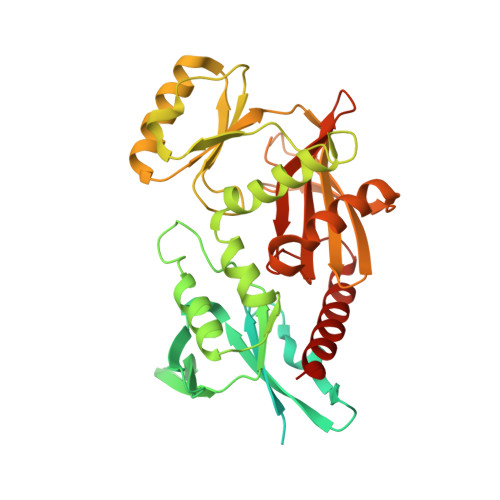
1PK8

Crystal Structure of Rat Synapsin I C Domain Complexed to Ca.ATP


Experimental Data Snapshot

  • Method: X-RAY DIFFRACTION
  • Resolution: 2.10 Å
  • R-Value Free: 0.259 
  • R-Value Work: 0.222 
  • R-Value Observed: 0.222 

wwPDB Validation   3D Report Full Report


Ligand Structure Quality Assessment 


This is version 1.4 of the entry. See complete history


Literature

Tetramerization and ATP binding by a protein comprising the A, B, and C domains of rat synapsin I.

Brautigam, C.A.Chelliah, Y.Deisenhofer, J.

(2004) J Biol Chem 279: 11948-11956

  • DOI: https://doi.org/10.1074/jbc.M312015200
  • Primary Citation of Related Structures:  
    1PK8, 1PX2

  • PubMed Abstract: 

    Synapsins are multidomain proteins that are critical for regulating neurotransmitter release in vertebrates. In the present study, two crystal structures of the C domain of rat synapsin I (rSynI-C) in complex with Ca(2+) and ATP reveal that this protein can form a tetramer and that a flexible loop (the "multifunctional loop") contacts bound ATP. Further experiments were carried out on a protein comprising the A, B, and C domains of rat synapsin I (rSynI-ABC). An ATP-stabilized tetramer of rSynI-ABC is observed during velocity sedimentation and size-exclusion chromatographic experiments. These hydrodynamic results also indicate that the A and B domains exist in an extended conformation. Calorimetric measurements of ATP binding to wild-type and mutant rSynI-ABC demonstrate that the multifunctional loop and a cross-tetramer contact are important for ATP binding. The evidence supports a view of synapsin I as an ATP-utilizing, tetrameric protein made up of monomers that have a flexible, extended N terminus.


  • Organizational Affiliation

    Howard Hughes Medical Institute and the Department of Biochemistry, The University of Texas Southwestern Medical Center at Dallas, Dallas, Texas 75390-9050, USA.


Macromolecules
Find similar proteins by:  (by identity cutoff)  |  3D Structure
Entity ID: 1
MoleculeChains Sequence LengthOrganismDetailsImage
rat synapsin I
A, B, C, D, E
A, B, C, D, E, F, G, H
422Rattus norvegicusMutation(s): 0 
Gene Names: SYN1
UniProt
Find proteins for P09951 (Rattus norvegicus)
Explore P09951 
Go to UniProtKB:  P09951
Entity Groups  
Sequence Clusters30% Identity50% Identity70% Identity90% Identity95% Identity100% Identity
UniProt GroupP09951
Sequence Annotations
Expand
  • Reference Sequence
Small Molecules
Ligands 3 Unique
IDChains Name / Formula / InChI Key2D Diagram3D Interactions
ATP
Query on ATP

Download Ideal Coordinates CCD File 
J [auth A]
M [auth B]
P [auth C]
R [auth D]
T [auth E]
J [auth A],
M [auth B],
P [auth C],
R [auth D],
T [auth E],
V [auth F],
X [auth G],
Z [auth H]
ADENOSINE-5'-TRIPHOSPHATE
C10 H16 N5 O13 P3
ZKHQWZAMYRWXGA-KQYNXXCUSA-N
EDO
Query on EDO

Download Ideal Coordinates CCD File 
K [auth A],
N [auth B]
1,2-ETHANEDIOL
C2 H6 O2
LYCAIKOWRPUZTN-UHFFFAOYSA-N
CA
Query on CA

Download Ideal Coordinates CCD File 
I [auth A]
L [auth B]
O [auth C]
Q [auth D]
S [auth E]
I [auth A],
L [auth B],
O [auth C],
Q [auth D],
S [auth E],
U [auth F],
W [auth G],
Y [auth H]
CALCIUM ION
Ca
BHPQYMZQTOCNFJ-UHFFFAOYSA-N
Binding Affinity Annotations 
IDSourceBinding Affinity
ATP Binding MOAD:  1PK8 Kd: 120 (nM) from 1 assay(s)
Experimental Data & Validation

Experimental Data

  • Method: X-RAY DIFFRACTION
  • Resolution: 2.10 Å
  • R-Value Free: 0.259 
  • R-Value Work: 0.222 
  • R-Value Observed: 0.222 
  • Space Group: P 1
Unit Cell:
Length ( Å )Angle ( ˚ )
a = 70.6α = 80.6
b = 78.4β = 76.9
c = 135γ = 71.8
Software Package:
Software NamePurpose
HKL-2000data collection
SCALEPACKdata scaling
EPMRphasing
CNSrefinement
HKL-2000data reduction

Structure Validation

View Full Validation Report


Ligand Structure Quality Assessment 


Entry History 

Deposition Data

Revision History  (Full details and data files)

  • Version 1.0: 2004-03-23
    Type: Initial release
  • Version 1.1: 2008-04-29
    Changes: Version format compliance
  • Version 1.2: 2011-07-13
    Changes: Version format compliance
  • Version 1.3: 2023-08-16
    Changes: Data collection, Database references, Derived calculations, Refinement description
  • Version 1.4: 2024-03-13
    Changes: Source and taxonomy, Structure summary