1PJR

STRUCTURE OF DNA HELICASE


Experimental Data Snapshot

  • Method: X-RAY DIFFRACTION
  • Resolution: 2.50 Å
  • R-Value Free: 0.269 
  • R-Value Work: 0.215 
  • R-Value Observed: 0.215 

wwPDB Validation   3D Report Full Report


This is version 1.3 of the entry. See complete history


Literature

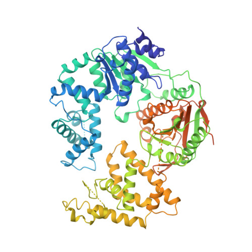
Crystal structure of a DExx box DNA helicase.

Subramanya, H.S.Bird, L.E.Brannigan, J.A.Wigley, D.B.

(1996) Nature 384: 379-383

  • DOI: https://doi.org/10.1038/384379a0
  • Primary Citation of Related Structures:  
    1PJR

  • PubMed Abstract: 

    There are a wide variety of helicases that unwind helical DNA and RNA substrates. The twelve helicases that have been identified in Escherichia coli play a role in almost all cellular processes involving nucleic acids. We have solved the crystal structure of a monomeric form of a DNA helicase from Bacillus stearothermophilus, alone and in a complex with ADP, at 2.5 and 2.9 A resolution, respectively. The enzyme comprises two domains with a deep cleft running between them. The ATP-binding site, which is situated at the bottom of this cleft, is formed by motifs that are conserved across the superfamily of related helicases. Unexpected structural homology with the DNA recombination protein, RecA, suggests how ATP binding and hydrolysis may drive conformational changes of the enzyme during catalysis, and implies that there is a common mechanism for all helicases.


  • Organizational Affiliation

    Laboratory of Molecular Biophysics, University of Oxford, UK.


Macromolecules
Find similar proteins by:  (by identity cutoff)  |  3D Structure
Entity ID: 1
MoleculeChains Sequence LengthOrganismDetailsImage
PCRA724Geobacillus stearothermophilusMutation(s): 0 
Gene Names: PCRA
UniProt
Find proteins for P56255 (Geobacillus stearothermophilus)
Explore P56255 
Go to UniProtKB:  P56255
Entity Groups  
Sequence Clusters30% Identity50% Identity70% Identity90% Identity95% Identity100% Identity
UniProt GroupP56255
Sequence Annotations
Expand
  • Reference Sequence
Experimental Data & Validation

Experimental Data

  • Method: X-RAY DIFFRACTION
  • Resolution: 2.50 Å
  • R-Value Free: 0.269 
  • R-Value Work: 0.215 
  • R-Value Observed: 0.215 
  • Space Group: P 65
Unit Cell:
Length ( Å )Angle ( ˚ )
a = 139.06α = 90
b = 139.06β = 90
c = 111.74γ = 120
Software Package:
Software NamePurpose
X-PLORmodel building
X-PLORrefinement
DENZOdata reduction
X-PLORphasing

Structure Validation

View Full Validation Report


Entry History 

Deposition Data

Revision History  (Full details and data files)

  • Version 1.0: 1997-12-03
    Type: Initial release
  • Version 1.1: 2008-03-24
    Changes: Version format compliance
  • Version 1.2: 2011-07-13
    Changes: Version format compliance
  • Version 1.3: 2024-02-14
    Changes: Data collection, Database references, Other