1PGF

PROSTAGLANDIN H2 SYNTHASE-1 COMPLEXED WITH 1-(4-IODOBENZOYL)-5-METHOXY-2-METHYLINDOLE-3-ACETIC ACID (IODOINDOMETHACIN), CIS MODEL


Experimental Data Snapshot

  • Method: X-RAY DIFFRACTION
  • Resolution: 4.50 Å
  • R-Value Free: 0.267 
  • R-Value Work: 0.254 
  • R-Value Observed: 0.254 

wwPDB Validation   3D Report Full Report


Ligand Structure Quality Assessment 


This is version 2.0 of the entry. See complete history


Literature

Synthesis and use of iodinated nonsteroidal antiinflammatory drug analogs as crystallographic probes of the prostaglandin H2 synthase cyclooxygenase active site.

Loll, P.J.Picot, D.Ekabo, O.Garavito, R.M.

(1996) Biochemistry 35: 7330-7340

  • DOI: https://doi.org/10.1021/bi952776w
  • Primary Citation of Related Structures:  
    1PGE, 1PGF, 1PGG

  • PubMed Abstract: 

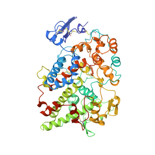
    The cyclooxygenase activity of the membrane protein prostaglandin H2 synthase isoform 1 (PGHS-1) is the target of the nonsteroidal antiinflammatory drugs (NSAIDs). The X-ray crystal structures of PGHS-1 in complex with the NSAIDs flurbiprofen and bromoaspirin have been determined previously [Picot, D., et al. (1994) Nature 367, 243-249; Loll, P. J., et al. (1995) Nat. Struct. Biol. 2, 637-643]. We report here the preparation and characterization of novel potent iodinated analogs of the NSAIDs indomethacin and suprofen, as well as the refined X-ray crystal structures of their complexes with PGHS-1. The PGHS-iodosuprofen complex structure has been refined at 3.5 A to an R-value of 0.189 and shows the suprofen analog to share a common mode of binding with flurbiprofen. The PGHS-iodoindomethacin complex structure has been refined at 4.5 A to an R-value of 0.254. The low resolution of the iodoindomethacin complex structure precludes detailed modeling of drug-enzyme interactions, but the electron-dense iodine atom of the inhibitor has been unambiguously located, allowing for the placement and approximate orientation of the inhibitor in the enzyme's active site. We have modeled two equally likely binding modes for iodoindomethacin, corresponding to the two principal conformers of the inhibitor. Like flurbiprofen, iodosuprofen and iodoindomethacin bind at the end of the long channel which leads into the enzyme active site. Binding at this site presumably blocks access of substrate to Tyr-385, a residue essential for catalysis. No evidence is seen for significant protein conformational differences between the iodoindomethacin and iodosuprofen of flurbiprofen complex structures.


  • Organizational Affiliation

    Department of Pharmacology, University of Pennsylvania School of Medicine, Philadelphia 19104-6084, USA.


Macromolecules
Find similar proteins by:  (by identity cutoff)  |  3D Structure
Entity ID: 1
MoleculeChains Sequence LengthOrganismDetailsImage
PROSTAGLANDIN H2 SYNTHASE-1
A, B
576Ovis ariesMutation(s): 0 
EC: 1.14.99.1
UniProt
Find proteins for P05979 (Ovis aries)
Explore P05979 
Go to UniProtKB:  P05979
Entity Groups  
Sequence Clusters30% Identity50% Identity70% Identity90% Identity95% Identity100% Identity
UniProt GroupP05979
Sequence Annotations
Expand
  • Reference Sequence
Oligosaccharides

Help

Entity ID: 2
MoleculeChains Length2D Diagram Glycosylation3D Interactions
2-acetamido-2-deoxy-beta-D-glucopyranose-(1-4)-2-acetamido-2-deoxy-beta-D-glucopyranose
C, D
2N-Glycosylation
Glycosylation Resources
GlyTouCan:  G42666HT
GlyCosmos:  G42666HT
GlyGen:  G42666HT
Small Molecules
Ligands 3 Unique
IDChains Name / Formula / InChI Key2D Diagram3D Interactions
HEM
Query on HEM

Download Ideal Coordinates CCD File 
G [auth A],
K [auth B]
PROTOPORPHYRIN IX CONTAINING FE
C34 H32 Fe N4 O4
KABFMIBPWCXCRK-RGGAHWMASA-L
IMM
Query on IMM

Download Ideal Coordinates CCD File 
H [auth A],
L [auth B]
1-(4-IODOBENZOYL)-5-METHOXY-2-METHYL INDOLE-3-ACETIC ACID
C19 H16 I N O4
CXBFZYKAVCAPSV-UHFFFAOYSA-N
NAG
Query on NAG

Download Ideal Coordinates CCD File 
E [auth A],
F [auth A],
I [auth B],
J [auth B]
2-acetamido-2-deoxy-beta-D-glucopyranose
C8 H15 N O6
OVRNDRQMDRJTHS-FMDGEEDCSA-N
Binding Affinity Annotations 
IDSourceBinding Affinity
IMM BindingDB:  1PGF IC50: 780 (nM) from 1 assay(s)
Experimental Data & Validation

Experimental Data

  • Method: X-RAY DIFFRACTION
  • Resolution: 4.50 Å
  • R-Value Free: 0.267 
  • R-Value Work: 0.254 
  • R-Value Observed: 0.254 
  • Space Group: I 2 2 2
Unit Cell:
Length ( Å )Angle ( ˚ )
a = 99.22α = 90
b = 208.99β = 90
c = 232.43γ = 90
Software Package:
Software NamePurpose
MADNESdata collection
X-PLORmodel building
X-PLORrefinement
MADNESdata reduction
X-PLORphasing

Structure Validation

View Full Validation Report


Ligand Structure Quality Assessment 


Entry History 

Deposition Data

Revision History  (Full details and data files)

  • Version 1.0: 1997-01-11
    Type: Initial release
  • Version 1.1: 2008-03-24
    Changes: Version format compliance
  • Version 1.2: 2011-07-13
    Changes: Non-polymer description, Version format compliance
  • Version 1.3: 2018-04-18
    Changes: Data collection, Other
  • Version 2.0: 2020-07-29
    Type: Remediation
    Reason: Carbohydrate remediation
    Changes: Atomic model, Data collection, Derived calculations, Structure summary