1PEY

Crystal structure of the Response Regulator Spo0F complexed with Mn2+


Experimental Data Snapshot

  • Method: X-RAY DIFFRACTION
  • Resolution: 2.25 Å
  • R-Value Free: 0.258 
  • R-Value Work: 0.225 
  • R-Value Observed: 0.225 

wwPDB Validation   3D Report Full Report


This is version 1.6 of the entry. See complete history


Literature

Metals in the sporulation phosphorelay: manganese binding by the response regulator Spo0F.

Mukhopadhyay, D.Sen, U.Zapf, J.Varughese, K.I.

(2004) Acta Crystallogr D Biol Crystallogr 60: 638-645

  • DOI: https://doi.org/10.1107/S0907444904002148
  • Primary Citation of Related Structures:  
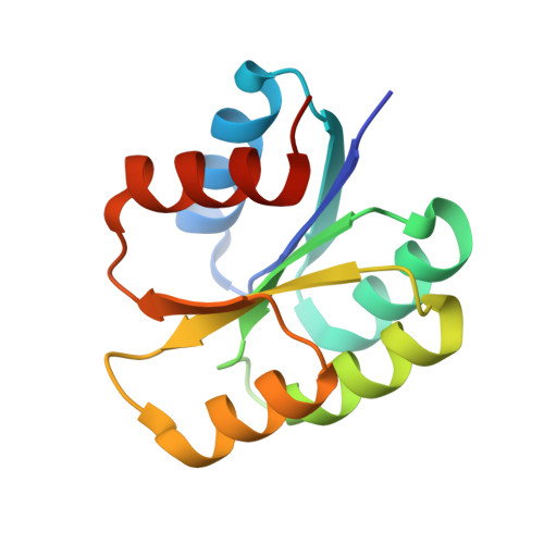
    1PEY

  • PubMed Abstract: 

    As a part of studies on the structural characterization of the components of the sporulation phosphorelay in Bacillus subtilis, the crystal structure of the manganese derivative of an intermediate signal transducer, Spo0F, has been elucidated at 2.25 A resolution. The calcium complex and the apo structures have been analyzed previously. In apo Spo0F, the active-site cation cavity is only partially formed and it only becomes completed upon metal coordination. The carbonyl of Lys56 is coordinated to the metal and interestingly the side chain of Lys56 exists in a variety of conformations in the three crystal structures of Spo0F. The affinity of the magnesium ion for Spo0F is in fact low; however, it binds Spo0F when it is in complex with Spo0B. It is proposed that the existence of a deep pocket which extends from the surface to the metal site could attract and direct the metal, thereby facilitating the metal binding of the complex.


  • Organizational Affiliation

    Division of Cellular Biology, Department of Molecular and Experimental Medicine, The Scripps Research Institute, La Jolla, CA 92037, USA.


Macromolecules
Find similar proteins by:  (by identity cutoff)  |  3D Structure
Entity ID: 1
MoleculeChains Sequence LengthOrganismDetailsImage
Sporulation initiation phosphotransferase F
A, B, C
124Bacillus subtilisMutation(s): 1 
Gene Names: SPO0F
EC: 2.7
UniProt
Find proteins for P06628 (Bacillus subtilis (strain 168))
Explore P06628 
Go to UniProtKB:  P06628
Entity Groups  
Sequence Clusters30% Identity50% Identity70% Identity90% Identity95% Identity100% Identity
UniProt GroupP06628
Sequence Annotations
Expand
  • Reference Sequence
Experimental Data & Validation

Experimental Data

  • Method: X-RAY DIFFRACTION
  • Resolution: 2.25 Å
  • R-Value Free: 0.258 
  • R-Value Work: 0.225 
  • R-Value Observed: 0.225 
  • Space Group: P 43 21 2
Unit Cell:
Length ( Å )Angle ( ˚ )
a = 103.128α = 90
b = 103.128β = 90
c = 83.641γ = 90
Software Package:
Software NamePurpose
DENZOdata reduction
SCALEPACKdata scaling
AMoREphasing
CNSrefinement

Structure Validation

View Full Validation Report


Entry History 

Deposition Data

Revision History  (Full details and data files)

  • Version 1.0: 2004-05-18
    Type: Initial release
  • Version 1.1: 2008-04-29
    Changes: Version format compliance
  • Version 1.2: 2011-07-13
    Changes: Version format compliance
  • Version 1.3: 2011-11-16
    Changes: Atomic model
  • Version 1.4: 2020-01-22
    Changes: Advisory, Derived calculations
  • Version 1.5: 2021-10-27
    Changes: Database references, Derived calculations
  • Version 1.6: 2024-02-14
    Changes: Data collection