1PBP

FINE TUNING OF THE SPECIFICITY OF THE PERIPLASMIC PHOSPHATE TRANSPORT RECEPTOR: SITE-DIRECTED MUTAGENESIS, LIGAND BINDING, AND CRYSTALLOGRAPHIC STUDIES


Experimental Data Snapshot

  • Method: X-RAY DIFFRACTION
  • Resolution: 1.90 Å
  • R-Value Work: 0.147 
  • R-Value Observed: 0.147 

wwPDB Validation   3D Report Full Report


This is version 1.3 of the entry. See complete history


Literature

Fine tuning the specificity of the periplasmic phosphate transport receptor. Site-directed mutagenesis, ligand binding, and crystallographic studies.

Wang, Z.Choudhary, A.Ledvina, P.S.Quiocho, F.A.

(1994) J Biol Chem 269: 25091-25094

  • DOI: https://doi.org/10.2210/pdb1pbp/pdb
  • Primary Citation of Related Structures:  
    1PBP

  • PubMed Abstract: 

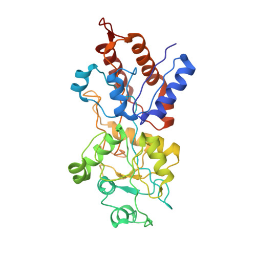
    Phosphorous, primarily in the form of phosphate, is a critical nutrient for the life of a cell. We have previously determined the 1.7-A resolution structure of the phosphate-binding protein, an initial receptor for the high-affinity phosphate active transport system or permease in Escherichia coli (Luecke, H., and Quiocho, F.A. (1990) Nature 347, 402-406). This structure is the first to reveal the key role of hydrogen bonding interactions in conferring the high specificity of the permease, a specificity also shared by other phosphate transport systems. Both monobasic and dibasic phosphates are recognized by the phosphate-binding protein with Asp56 playing a key role. Here we report site-directed mutagenesis, ligand binding, and crystallographic studies of the binding protein which show that introduction of one additional Asp by mutagenesis of the Thr141 in the ligand-binding site restricts binding to only the monobasic phosphate.


  • Organizational Affiliation

    Structural and Computational Biology and Molecular Biophysics Program, Baylor College of Medicine, Houston, Texas 77030.


Macromolecules
Find similar proteins by:  (by identity cutoff)  |  3D Structure
Entity ID: 1
MoleculeChains Sequence LengthOrganismDetailsImage
PHOSPHATE-BINDING PROTEIN321Escherichia coliMutation(s): 0 
UniProt
Find proteins for P0AG82 (Escherichia coli (strain K12))
Explore P0AG82 
Go to UniProtKB:  P0AG82
Entity Groups  
Sequence Clusters30% Identity50% Identity70% Identity90% Identity95% Identity100% Identity
UniProt GroupP0AG82
Sequence Annotations
Expand
  • Reference Sequence
Small Molecules
Ligands 1 Unique
IDChains Name / Formula / InChI Key2D Diagram3D Interactions
PO4
Query on PO4

Download Ideal Coordinates CCD File 
B [auth A]PHOSPHATE ION
O4 P
NBIIXXVUZAFLBC-UHFFFAOYSA-K
Experimental Data & Validation

Experimental Data

  • Method: X-RAY DIFFRACTION
  • Resolution: 1.90 Å
  • R-Value Work: 0.147 
  • R-Value Observed: 0.147 
  • Space Group: P 21 21 21
Unit Cell:
Length ( Å )Angle ( ˚ )
a = 41.88α = 90
b = 64.57β = 90
c = 124.67γ = 90
Software Package:
Software NamePurpose
X-PLORmodel building
X-PLORrefinement
X-PLORphasing

Structure Validation

View Full Validation Report


Entry History 

Deposition Data

Revision History  (Full details and data files)

  • Version 1.0: 1994-10-15
    Type: Initial release
  • Version 1.1: 2008-03-24
    Changes: Version format compliance
  • Version 1.2: 2011-07-13
    Changes: Version format compliance
  • Version 1.3: 2024-02-14
    Changes: Data collection, Database references, Derived calculations, Other