1PAQ

CRYSTAL STRUCTURE OF THE CATALYTIC FRAGMENT OF EUKARYOTIC INITIATION FACTOR 2B EPSILON


Experimental Data Snapshot

  • Method: X-RAY DIFFRACTION
  • Resolution: 2.30 Å
  • R-Value Free: 0.272 
  • R-Value Work: 0.243 
  • R-Value Observed: 0.244 

wwPDB Validation   3D Report Full Report


This is version 1.2 of the entry. See complete history


Literature

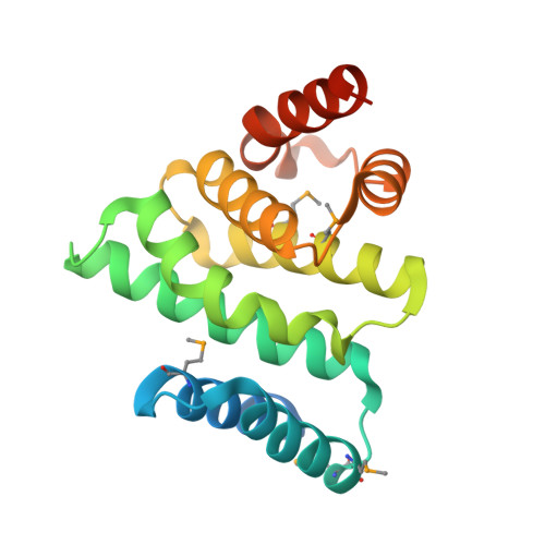
Structure of the catalytic fragment of translation initiation factor 2B and identification of a critically important catalytic residue.

Boesen, T.Mohammad, S.S.Pavitt, G.D.Andersen, G.R.

(2004) J Biol Chem 279: 10584-10592

  • DOI: https://doi.org/10.1074/jbc.M311055200
  • Primary Citation of Related Structures:  
    1PAQ

  • PubMed Abstract: 

    Eukaryotic initiation factor (eIF) 2B catalyzes the nucleotide activation of eIF2 to its active GTP-bound state. The exchange activity has been mapped to the C terminus of the eIF2Bepsilon subunit. We have determined the crystal structure of residues 544-704 from yeast eIF2Bepsilon at 2.3-A resolution, and this fragment is an all-helical protein built around the conserved aromatic acidic (AA) boxes also found in eIF4G and eIF5. The eight helices are organized in a manner similar to HEAT repeats. The molecule is highly asymmetric with respect to surface charge and conservation. One area in the N terminus is proposed to be directly involved in catalysis. In agreement with this hypothesis, mutation of glutamate 569 is shown to be lethal. An acidic belt and a second area in the C terminus containing residues from the AA boxes are important for binding to eIF2. Two mutations causing the fatal human genetic disease leukoencephalopathy with vanishing white matter are buried and appear to disrupt the structural integrity of the catalytic domain rather than interfering directly with catalysis or binding of eIF2.


  • Organizational Affiliation

    Department of Molecular Biology, Aarhus University, Denmark.


Macromolecules
Find similar proteins by:  (by identity cutoff)  |  3D Structure
Entity ID: 1
MoleculeChains Sequence LengthOrganismDetailsImage
Translation initiation factor eIF-2B epsilon subunit189Saccharomyces cerevisiaeMutation(s): 5 
Gene Names: GCD6 OR TIF225 OR YDR211W OR YD8142.12 OR YD8142B.03
UniProt
Find proteins for P32501 (Saccharomyces cerevisiae (strain ATCC 204508 / S288c))
Explore P32501 
Go to UniProtKB:  P32501
Entity Groups  
Sequence Clusters30% Identity50% Identity70% Identity90% Identity95% Identity100% Identity
UniProt GroupP32501
Sequence Annotations
Expand
  • Reference Sequence
Small Molecules
Modified Residues  1 Unique
IDChains TypeFormula2D DiagramParent
MSE
Query on MSE
A
L-PEPTIDE LINKINGC5 H11 N O2 SeMET
Experimental Data & Validation

Experimental Data

  • Method: X-RAY DIFFRACTION
  • Resolution: 2.30 Å
  • R-Value Free: 0.272 
  • R-Value Work: 0.243 
  • R-Value Observed: 0.244 
  • Space Group: I 4 2 2
Unit Cell:
Length ( Å )Angle ( ˚ )
a = 106.34α = 90
b = 106.34β = 90
c = 91.71γ = 90
Software Package:
Software NamePurpose
MOSFLMdata reduction
SCALAdata scaling
CNSrefinement
CCP4data scaling
CNSphasing

Structure Validation

View Full Validation Report


Entry History 

Deposition Data

Revision History  (Full details and data files)

  • Version 1.0: 2004-02-10
    Type: Initial release
  • Version 1.1: 2008-04-29
    Changes: Version format compliance
  • Version 1.2: 2011-07-13
    Changes: Version format compliance