1P7G

Crystal structure of superoxide dismutase from Pyrobaculum aerophilum


Experimental Data Snapshot

  • Method: X-RAY DIFFRACTION
  • Resolution: 1.80 Å
  • R-Value Free: 0.217 
  • R-Value Work: 0.162 
  • R-Value Observed: 0.162 

wwPDB Validation   3D Report Full Report


This is version 1.4 of the entry. See complete history


Literature

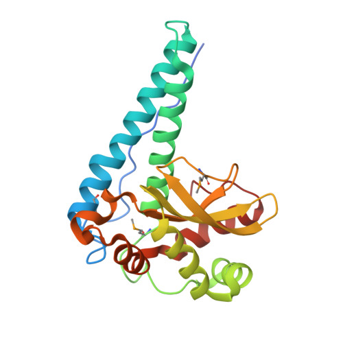
Structure of superoxide dismutase from Pyrobaculum aerophilum presents a challenging case in molecular replacement with multiple molecules, pseudo-symmetry and twinning.

Lee, S.Sawaya, M.R.Eisenberg, D.

(2003) Acta Crystallogr D Biol Crystallogr 59: 2191-2199

  • DOI: https://doi.org/10.1107/s0907444903019942
  • Primary Citation of Related Structures:  
    1P7G

  • PubMed Abstract: 

    The crystal structure of superoxide dismutase from the hyperthermophilic crenarchaeon Pyrobaculum aerophilum was determined by molecular replacement at 1.8 A resolution. The structure determination was made especially challenging by the large number of molecules (24) in the asymmetric unit, the presence of a pseudo-crystallographic twofold operator close to a twinning operator and the inability to detect twinning by conventional means. Molecular replacement proceeded at low resolution in pseudo (apparent) space group P3(2)12 and was facilitated by examination of the self-rotation function and native Patterson map. Refinement, however, stalled at an R factor of 40% when high-resolution data were included. Expanding to the lower symmetry space group P3(2) decreased R (to 22%) and R(free) (to 26%), but not by as much as expected for the quality of data. Finally, despite the apparent lack of evidence from conventional twinning tests [i.e. plots of the second moment of I and N(Z) distributions], a twinning operator was included in the refinement, lowering R and R(free) to 16.2 and 21.7%, respectively. The early detection of twinning appears to have been masked by a deviation in the expected intensity distribution caused by the presence of non-crystallographic translational symmetry. These findings suggest the importance of testing twinning operators in cases where pseudo-translational symmetry can explain negative results from conventional twinning tests. The structure reveals a tetrameric assembly with 222 symmetry, similar to superoxide dismutase structures from other organisms. The current structural model represents the metal-free state of the enzyme.


  • Organizational Affiliation

    UCLA-DOE Institute for Genomics and Proteomics, Molecular Biology Institute, Howard Hughes Medical Institute, and Department of Chemistry and Biochemistry, University of California,Los Angeles, Los Angeles, CA 90095-1570, USA.


Macromolecules
Find similar proteins by:  (by identity cutoff)  |  3D Structure
Entity ID: 1
MoleculeChains Sequence LengthOrganismDetailsImage
Superoxide dismutase
A, B, C, D, E
A, B, C, D, E, F, G, H, I, J, K, L, M, N, O, P, Q, R, S, T, U, V, W, X
222Pyrobaculum aerophilumMutation(s): 3 
Gene Names: SOD OR PAE0274
EC: 1.15.1.1
UniProt
Find proteins for O93724 (Pyrobaculum aerophilum (strain ATCC 51768 / DSM 7523 / JCM 9630 / CIP 104966 / NBRC 100827 / IM2))
Explore O93724 
Go to UniProtKB:  O93724
Entity Groups  
Sequence Clusters30% Identity50% Identity70% Identity90% Identity95% Identity100% Identity
UniProt GroupO93724
Sequence Annotations
Expand
  • Reference Sequence
Small Molecules
Ligands 2 Unique
IDChains Name / Formula / InChI Key2D Diagram3D Interactions
BME
Query on BME

Download Ideal Coordinates CCD File 
BA [auth D]
CB [auth X]
EA [auth F]
GA [auth H]
HA [auth I]
BA [auth D],
CB [auth X],
EA [auth F],
GA [auth H],
HA [auth I],
JA [auth K],
NA [auth N],
QA [auth P],
SA [auth Q],
WA [auth T],
Z [auth B],
ZA [auth V]
BETA-MERCAPTOETHANOL
C2 H6 O S
DGVVWUTYPXICAM-UHFFFAOYSA-N
ACT
Query on ACT

Download Ideal Coordinates CCD File 
AA [auth D]
AB [auth W]
BB [auth X]
CA [auth E]
DA [auth F]
AA [auth D],
AB [auth W],
BB [auth X],
CA [auth E],
DA [auth F],
FA [auth H],
IA [auth J],
KA [auth L],
LA [auth M],
MA [auth N],
OA [auth O],
PA [auth P],
RA [auth Q],
TA [auth R],
UA [auth S],
VA [auth T],
XA [auth U],
Y [auth B],
YA [auth V]
ACETATE ION
C2 H3 O2
QTBSBXVTEAMEQO-UHFFFAOYSA-M
Modified Residues  1 Unique
IDChains TypeFormula2D DiagramParent
MSE
Query on MSE
A, B, C, D, E
A, B, C, D, E, F, G, H, I, J, K, L, M, N, O, P, Q, R, S, T, U, V, W, X
L-PEPTIDE LINKINGC5 H11 N O2 SeMET
Experimental Data & Validation

Experimental Data

  • Method: X-RAY DIFFRACTION
  • Resolution: 1.80 Å
  • R-Value Free: 0.217 
  • R-Value Work: 0.162 
  • R-Value Observed: 0.162 
  • Space Group: P 32
Unit Cell:
Length ( Å )Angle ( ˚ )
a = 163.429α = 90
b = 163.429β = 90
c = 172.169γ = 120
Software Package:
Software NamePurpose
DENZOdata reduction
SCALEPACKdata scaling
GLRFphasing
CNSrefinement

Structure Validation

View Full Validation Report


Entry History 

Deposition Data

Revision History  (Full details and data files)

  • Version 1.0: 2003-12-02
    Type: Initial release
  • Version 1.1: 2008-04-29
    Changes: Version format compliance
  • Version 1.2: 2011-07-13
    Changes: Version format compliance
  • Version 1.3: 2023-08-16
    Changes: Data collection, Database references, Derived calculations, Refinement description
  • Version 1.4: 2023-11-15
    Changes: Data collection