1P6C

crystal structure of phosphotriesterase triple mutant H254G/H257W/L303T complexed with diisopropylmethylphosphonate


Experimental Data Snapshot

  • Method: X-RAY DIFFRACTION
  • Resolution: 2.00 Å
  • R-Value Free: 0.246 
  • R-Value Work: 0.181 
  • R-Value Observed: 0.185 

wwPDB Validation   3D Report Full Report


Ligand Structure Quality Assessment 


This is version 1.6 of the entry. See complete history


Literature

Enhanced degradation of chemical warfare agents through molecular engineering of the phosphotriesterase active site.

Hill, C.M.Li, W.S.Thoden, J.B.Holden, H.M.Raushel, F.M.

(2003) J Am Chem Soc 125: 8990-8991

  • DOI: https://doi.org/10.1021/ja0358798
  • Primary Citation of Related Structures:  
    1P6B, 1P6C

  • PubMed Abstract: 

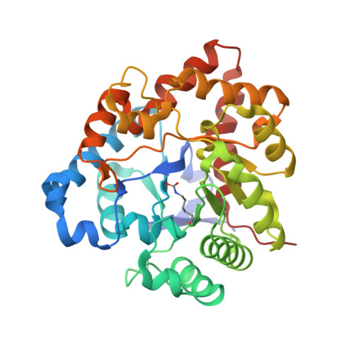
    The bacterial phosphotriesterase has been utilized as a template for the evolution of improved enzymes for the catalytic decomposition of organophosphate nerve agents. A combinatorial library of active site mutants was constructed by randomizing residues His-254, His-257, and Leu-303. The collection of mutant proteins was screened for the ability to hydrolyze a chromogenic analogue of the most toxic stereoisomer of the chemical warfare agent, soman. The mutant H254G/H257W/L303T catalyzed the hydrolysis of the target substrate nearly 3 orders of magnitude faster than the wild-type enzyme. The X-ray crystal structure was solved in the presence and absence of diisopropyl methyl phosphonate. The mutant enzyme was ligated to an additional divalent cation at the active site that was displaced upon the binding of the substrate analogue inhibitor. These studies demonstrate that substantial changes in substrate specificity can be achieved by relatively minor changes to the primary amino acid sequence.


  • Organizational Affiliation

    Department of Chemistry, P.O. Box 30012, Texas A&M University, College Station, TX 77842-3012, USA.


Macromolecules
Find similar proteins by:  (by identity cutoff)  |  3D Structure
Entity ID: 1
MoleculeChains Sequence LengthOrganismDetailsImage
Parathion hydrolase
A, B
336Flavobacterium sp.Mutation(s): 3 
Gene Names: OPD
EC: 3.1.8.1
UniProt
Find proteins for P0A433 (Sphingobium fuliginis (strain ATCC 27551))
Explore P0A433 
Go to UniProtKB:  P0A433
Entity Groups  
Sequence Clusters30% Identity50% Identity70% Identity90% Identity95% Identity100% Identity
UniProt GroupP0A433
Sequence Annotations
Expand
  • Reference Sequence
Small Molecules
Ligands 3 Unique
IDChains Name / Formula / InChI Key2D Diagram3D Interactions
EBP
Query on EBP

Download Ideal Coordinates CCD File 
E [auth A],
I [auth B]
DIETHYL 4-METHYLBENZYLPHOSPHONATE
C12 H19 O3 P
QKGBKPZAXXBLJE-UHFFFAOYSA-N
DII
Query on DII

Download Ideal Coordinates CCD File 
F [auth A],
J [auth B]
METHYLPHOSPHONIC ACID DIISOPROPYL ESTER
C7 H17 O3 P
WOAFDHWYKSOANX-UHFFFAOYSA-N
ZN
Query on ZN

Download Ideal Coordinates CCD File 
C [auth A],
D [auth A],
G [auth B],
H [auth B]
ZINC ION
Zn
PTFCDOFLOPIGGS-UHFFFAOYSA-N
Modified Residues  1 Unique
IDChains TypeFormula2D DiagramParent
KCX
Query on KCX
A, B
L-PEPTIDE LINKINGC7 H14 N2 O4LYS
Experimental Data & Validation

Experimental Data

  • Method: X-RAY DIFFRACTION
  • Resolution: 2.00 Å
  • R-Value Free: 0.246 
  • R-Value Work: 0.181 
  • R-Value Observed: 0.185 
  • Space Group: C 1 2 1
Unit Cell:
Length ( Å )Angle ( ˚ )
a = 130.4α = 90
b = 91.9β = 90.9
c = 69.7γ = 90
Software Package:
Software NamePurpose
FRAMBOdata collection
SAINTdata reduction
TNTrefinement
SAINTdata scaling
TNTphasing

Structure Validation

View Full Validation Report


Ligand Structure Quality Assessment 


Entry History 

Deposition Data

Revision History  (Full details and data files)

  • Version 1.0: 2003-09-16
    Type: Initial release
  • Version 1.1: 2008-04-29
    Changes: Version format compliance
  • Version 1.2: 2011-07-13
    Changes: Version format compliance
  • Version 1.3: 2017-10-11
    Changes: Refinement description
  • Version 1.4: 2021-10-27
    Changes: Database references, Derived calculations
  • Version 1.5: 2023-08-16
    Changes: Data collection, Refinement description
  • Version 1.6: 2023-11-15
    Changes: Data collection