1P5E

The structure of phospho-CDK2/cyclin A in complex with the inhibitor 4,5,6,7-tetrabromobenzotriazole (TBS)


Experimental Data Snapshot

  • Method: X-RAY DIFFRACTION
  • Resolution: 2.22 Å
  • R-Value Free: 0.257 
  • R-Value Work: 0.219 
  • R-Value Observed: 0.219 

wwPDB Validation   3D Report Full Report


Ligand Structure Quality Assessment 


This is version 1.3 of the entry. See complete history


Literature

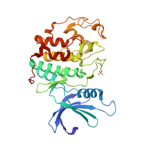
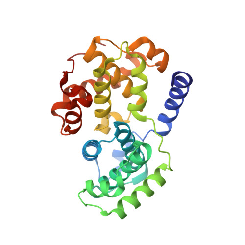
Alternative binding modes of an inhibitor to two different kinases

De Moliner, E.Brown, N.R.Johnson, L.N.

(2003) Eur J Biochem 270: 1-8

  • DOI: https://doi.org/10.1046/j.1432-1033.2003.03697.x
  • Primary Citation of Related Structures:  
    1P5E

  • PubMed Abstract: 

    Protein kinases are targets for therapeutic agents designed to intervene in signaling processes in the diseased state. Most kinase inhibitors are directed towards the conserved ATP binding site. Because the essential features of this site are conserved in all eukaryotic protein kinases, it is generally assumed that the same compound will bind in a similar manner to different protein kinases. The inhibitor 4,5,6,7-tetrabromobenzotriazole (TBB) is a selective inhibitor for the protein kinase CK2 (IC50 1.6 micro m) (Sarno et al. (2001) FEBS Letts.496, 44-48). Three other kinases [cyclin-dependent protein kinase 2 (CDK2), phosphorylase kinase and glycogen synthase kinase 3beta] exhibit approximately 10-fold weaker affinity for TBB than CK2. We report the crystal structure of TBB in complex with phospho-CDK2-cyclin A at 2.2 A resolution and compare the interactions with those observed for TBB bound to CK2. TBB binds at the ATP binding site of both kinases. In CDK2, each of the four bromine atoms makes polar contacts either to main chain oxygens in the hinge region of the kinase or to water molecules, in addition to several van der Waals contacts. The mode of binding of TBB to CDK2 is different from that to CK2. TBB in CDK2 is displaced more towards the hinge region between the N- and C-terminal lobes and rotated relative to TBB in CK2. The ATP binding pocket is wider in CDK2 than in CK2 resulting in fewer van der Waals contacts but TBB in CK2 does not contact the hinge. The structures show that, despite the conservation of the ATP binding pocket, the inhibitor is able to exploit different recognition features so that the same compound can bind in different ways to the two different kinases.


  • Organizational Affiliation

    Laboratory of Molecular Biophysics, Department of Biochemistry, University of Oxford, UK.


Macromolecules
Find similar proteins by:  (by identity cutoff)  |  3D Structure
Entity ID: 1
MoleculeChains Sequence LengthOrganismDetailsImage
Cell division protein kinase 2
A, C
299Homo sapiensMutation(s): 1 
Gene Names: CDK2
EC: 2.7.1
UniProt & NIH Common Fund Data Resources
Find proteins for P24941 (Homo sapiens)
Explore P24941 
Go to UniProtKB:  P24941
PHAROS:  P24941
GTEx:  ENSG00000123374 
Entity Groups  
Sequence Clusters30% Identity50% Identity70% Identity90% Identity95% Identity100% Identity
UniProt GroupP24941
Sequence Annotations
Expand
  • Reference Sequence
Find similar proteins by:  (by identity cutoff)  |  3D Structure
Entity ID: 2
MoleculeChains Sequence LengthOrganismDetailsImage
Cyclin A2
B, D
258Homo sapiensMutation(s): 0 
Gene Names: CCNA2 OR CCNA OR CCN1
UniProt & NIH Common Fund Data Resources
Find proteins for P20248 (Homo sapiens)
Explore P20248 
Go to UniProtKB:  P20248
PHAROS:  P20248
GTEx:  ENSG00000145386 
Entity Groups  
Sequence Clusters30% Identity50% Identity70% Identity90% Identity95% Identity100% Identity
UniProt GroupP20248
Sequence Annotations
Expand
  • Reference Sequence
Small Molecules
Ligands 1 Unique
IDChains Name / Formula / InChI Key2D Diagram3D Interactions
TBS
Query on TBS

Download Ideal Coordinates CCD File 
E [auth A],
F [auth C]
4,5,6,7-TETRABROMOBENZOTRIAZOLE
C6 H Br4 N3
OMZYUVOATZSGJY-UHFFFAOYSA-N
Modified Residues  1 Unique
IDChains TypeFormula2D DiagramParent
TPO
Query on TPO
A, C
L-PEPTIDE LINKINGC4 H10 N O6 PTHR
Binding Affinity Annotations 
IDSourceBinding Affinity
TBS PDBBind:  1P5E IC50: 1.56e+4 (nM) from 1 assay(s)
Experimental Data & Validation

Experimental Data

  • Method: X-RAY DIFFRACTION
  • Resolution: 2.22 Å
  • R-Value Free: 0.257 
  • R-Value Work: 0.219 
  • R-Value Observed: 0.219 
  • Space Group: P 21 21 21
Unit Cell:
Length ( Å )Angle ( ˚ )
a = 73.536α = 90
b = 133.947β = 90
c = 148.424γ = 90
Software Package:
Software NamePurpose
MOSFLMdata reduction
SCALAdata scaling
CNSrefinement
CCP4data scaling
CNSphasing

Structure Validation

View Full Validation Report


Ligand Structure Quality Assessment 


Entry History 

Deposition Data

Revision History  (Full details and data files)

  • Version 1.0: 2003-07-01
    Type: Initial release
  • Version 1.1: 2008-04-29
    Changes: Version format compliance
  • Version 1.2: 2011-07-13
    Changes: Version format compliance
  • Version 1.3: 2016-12-21
    Changes: Structure summary