1P4U

CRYSTAL STRUCTURE OF GGA3 GAE DOMAIN IN COMPLEX WITH RABAPTIN-5 PEPTIDE


Experimental Data Snapshot

  • Method: X-RAY DIFFRACTION
  • Resolution: 2.20 Å
  • R-Value Free: 0.255 
  • R-Value Work: 0.217 
  • R-Value Observed: 0.217 

wwPDB Validation   3D Report Full Report


This is version 1.4 of the entry. See complete history


Literature

RECOGNITION OF ACCESSORY PROTEIN MOTIFS BY THE GAMMA-ADAPTIN EAR DOMAIN OF GGA3

MILLER, G.J.MATTERA, R.BONIFACINO, J.S.HURLEY, J.H.

(2003) Nat Struct Biol 10: 599-606

  • DOI: https://doi.org/10.1038/nsb953
  • Primary Citation of Related Structures:  
    1P4U

  • PubMed Abstract: 

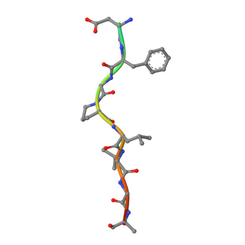
    Adaptor proteins load transmembrane protein cargo into transport vesicles and serve as nexuses for the formation of large multiprotein complexes on the nascent vesicles. The gamma-adaptin ear (GAE) domains of the AP-1 adaptor protein complex and the GGA adaptor proteins recruit accessory proteins to these multiprotein complexes by binding to a hydrophobic motif. We determined the structure of the GAE domain of human GGA3 in complex with a peptide based on the DFGPLV sequence of the accessory protein Rabaptin-5 and refined it at a resolution of 2.2 A. The leucine and valine residues of the peptide are partly buried in two contiguous shallow, hydrophobic depressions. The anchoring phenylalanine is buried in a deep pocket formed by the aliphatic portions of two conserved arginine residues, along with an alanine and a proline, illustrating the unusual function of a cluster of basic residues in binding a hydrophobic motif.


  • Organizational Affiliation

    Laboratory of Molecular Biology, National Institute of Diabetes and Digestive and Kidney Diseases, National Institutes of Health, Department of Health and Human Services, Bethesda, Maryland 20892, USA.


Macromolecules
Find similar proteins by:  (by identity cutoff)  |  3D Structure
Entity ID: 1
MoleculeChains Sequence LengthOrganismDetailsImage
ADP-ribosylation factor binding protein GGA3153Homo sapiensMutation(s): 2 
Gene Names: GGA3
Membrane Entity: Yes 
UniProt & NIH Common Fund Data Resources
Find proteins for Q9NZ52 (Homo sapiens)
Explore Q9NZ52 
Go to UniProtKB:  Q9NZ52
PHAROS:  Q9NZ52
GTEx:  ENSG00000125447 
Entity Groups  
Sequence Clusters30% Identity50% Identity70% Identity90% Identity95% Identity100% Identity
UniProt GroupQ9NZ52
Sequence Annotations
Expand
  • Reference Sequence

Find similar proteins by:  Sequence   |   3D Structure  

Entity ID: 2
MoleculeChains Sequence LengthOrganismDetailsImage
Rabaptin-513N/AMutation(s): 0 
UniProt & NIH Common Fund Data Resources
Find proteins for Q15276 (Homo sapiens)
Explore Q15276 
Go to UniProtKB:  Q15276
PHAROS:  Q15276
GTEx:  ENSG00000029725 
Entity Groups  
Sequence Clusters30% Identity50% Identity70% Identity90% Identity95% Identity100% Identity
UniProt GroupQ15276
Sequence Annotations
Expand
  • Reference Sequence
Experimental Data & Validation

Experimental Data

  • Method: X-RAY DIFFRACTION
  • Resolution: 2.20 Å
  • R-Value Free: 0.255 
  • R-Value Work: 0.217 
  • R-Value Observed: 0.217 
  • Space Group: C 2 2 21
Unit Cell:
Length ( Å )Angle ( ˚ )
a = 45.2α = 90
b = 85.2β = 90
c = 97γ = 90
Software Package:
Software NamePurpose
CNSrefinement
DENZOdata reduction
SCALEPACKdata scaling

Structure Validation

View Full Validation Report


Entry History 

Deposition Data

Revision History  (Full details and data files)

  • Version 1.0: 2003-07-29
    Type: Initial release
  • Version 1.1: 2008-04-29
    Changes: Version format compliance
  • Version 1.2: 2011-07-13
    Changes: Version format compliance
  • Version 1.3: 2021-10-27
    Changes: Database references
  • Version 1.4: 2024-02-14
    Changes: Data collection