High Resolution Structures of an Oxidized and Reduced Flavoprotein: THE WATER SWITCH IN A SOLUBLE FORM OF (S)-MANDELATE DEHYDROGENASE
Sukumar, N., Dewanti, A.R., Mitra, B., Mathews, F.S.(2004) J Biol Chem 279: 3749-3757
- PubMed: 14604988
- DOI: https://doi.org/10.1074/jbc.M310049200
- Primary Citation of Related Structures:
1P4C, 1P5B - PubMed Abstract:
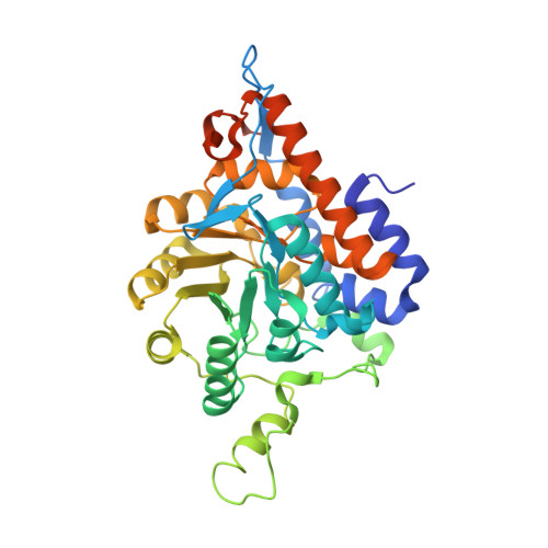
The crystal structures of a soluble mutant of the flavoenzyme mandelate dehydrogenase (MDH) from Pseudomonas putida and of the substrate-reduced enzyme have been analyzed at 1.35-A resolution. The mutant (MDH-GOX2) is a fully active chimeric enzyme in which residues 177-215 of the membrane-bound MDH are replaced by residues 176-195 of glycolate oxidase from spinach. Both structures permit full tracing of the polypeptide backbone chain from residues 4-356, including a 4-residue segment that was disordered in an earlier study of the oxidized protein at 2.15 A resolution. The structures of MDH-GOX2 in the oxidized and reduced states are virtually identical with only a slight increase in the bending angle of the flavin ring upon reduction. The only other structural changes within the protein interior are a 10 degrees rotation of an active site tyrosine side chain, the loss of an active site water, and a significant movement of six other water molecules in the active site by 0.45 to 0.78 A. Consistent with solution studies, there is no apparent binding of either the substrate, mandelate, or the oxidation product, benzoylformate, to the reduced enzyme. The observed structural changes upon enzyme reduction have been interpreted as a rearrangement of the hydrogen bonding pattern within the active site that results from binding of a proton to the N-5 position of the anionic hydroquinone form of the reduced flavin prosthetic group. Implications for the low oxidase activity of the reduced enzyme are also discussed.
Organizational Affiliation:
Department of Biochemistry and Molecular Biophysics, Washington University School of Medicine, St. Louis, Missouri 63110, USA.