1P3Q

Mechanism of Ubiquitin Recognition by the CUE Domain of VPS9


Experimental Data Snapshot

  • Method: X-RAY DIFFRACTION
  • Resolution: 1.70 Å
  • R-Value Free: 0.277 
  • R-Value Work: 0.260 

wwPDB Validation   3D Report Full Report


This is version 1.4 of the entry. See complete history


Literature

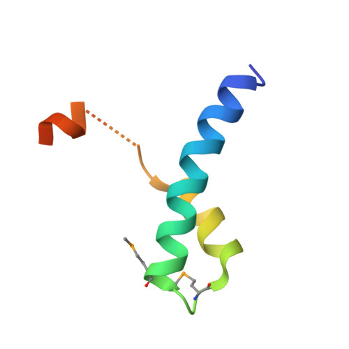
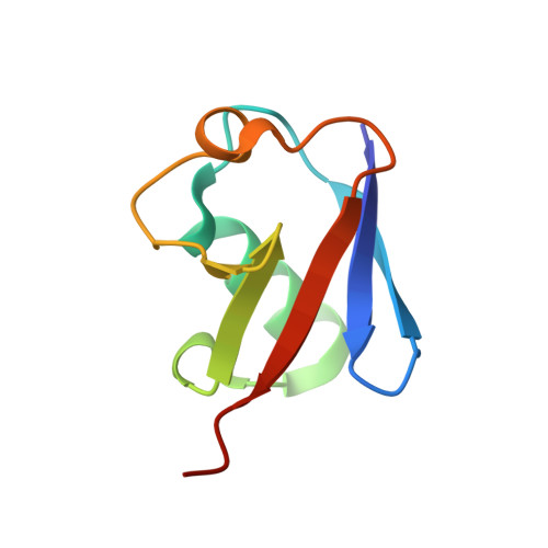
Mechanism of Ubiquitin Recognition by the CUE Domain of Vps9p.

Prag, G.Misra, S.Jones, E.A.Ghirlando, R.Davies, B.A.Horazdovsky, B.F.Hurley, J.H.

(2003) Cell 113: 609-620

  • DOI: https://doi.org/10.1016/s0092-8674(03)00364-7
  • Primary Citation of Related Structures:  
    1MN3, 1P3Q

  • PubMed Abstract: 

    Coupling of ubiquitin conjugation to ER degradation (CUE) domains are approximately 50 amino acid monoubiquitin binding motifs found in proteins of trafficking and ubiquitination pathways. The 2.3 A structure of the Vps9p-CUE domain is a dimeric domain-swapped variant of the ubiquitin binding UBA domain. The 1.7 A structure of the CUE:ubiquitin complex shows that one CUE dimer binds one ubiquitin molecule. The bound CUE dimer is kinked relative to the unbound CUE dimer and wraps around ubiquitin. The CUE monomer contains two ubiquitin binding surfaces on opposite faces of the molecule that cannot bind simultaneously to a single ubiquitin molecule. Dimerization of the CUE domain allows both surfaces to contact a single ubiquitin molecule, providing a mechanism for high-affinity binding to monoubiquitin.


  • Organizational Affiliation

    Laboratory of Molecular Biology, National Institute of Diabetes and Digestive and Kidney Diseases, National Institutes of Health, Department of Health and Human Services, Bethesda, MD 20892, USA.


Macromolecules
Find similar proteins by:  (by identity cutoff)  |  3D Structure
Entity ID: 1
MoleculeChains Sequence LengthOrganismDetailsImage
Vacuolar protein sorting-associated protein VPS9A [auth Q],
B [auth R]
54Saccharomyces cerevisiaeMutation(s): 2 
Gene Names: VPS9
UniProt
Find proteins for P54787 (Saccharomyces cerevisiae (strain ATCC 204508 / S288c))
Explore P54787 
Go to UniProtKB:  P54787
Entity Groups  
Sequence Clusters30% Identity50% Identity70% Identity90% Identity95% Identity100% Identity
UniProt GroupP54787
Sequence Annotations
Expand
  • Reference Sequence
Find similar proteins by:  (by identity cutoff)  |  3D Structure
Entity ID: 2
MoleculeChains Sequence LengthOrganismDetailsImage
UbiquitinC [auth U],
D [auth V]
76Bos taurusMutation(s): 0 
UniProt
Find proteins for P0CH28 (Bos taurus)
Explore P0CH28 
Go to UniProtKB:  P0CH28
Entity Groups  
Sequence Clusters30% Identity50% Identity70% Identity90% Identity95% Identity100% Identity
UniProt GroupP0CH28
Sequence Annotations
Expand
  • Reference Sequence
Small Molecules
Modified Residues  1 Unique
IDChains TypeFormula2D DiagramParent
MSE
Query on MSE
A [auth Q],
B [auth R]
L-PEPTIDE LINKINGC5 H11 N O2 SeMET
Experimental Data & Validation

Experimental Data

  • Method: X-RAY DIFFRACTION
  • Resolution: 1.70 Å
  • R-Value Free: 0.277 
  • R-Value Work: 0.260 
  • Space Group: C 1 2 1
Unit Cell:
Length ( Å )Angle ( ˚ )
a = 101.607α = 90
b = 45.887β = 96.53
c = 57.802γ = 90
Software Package:
Software NamePurpose
CNSrefinement
MAR345data collection
SCALEPACKdata scaling
CNSphasing

Structure Validation

View Full Validation Report


Entry History 

Deposition Data

Revision History  (Full details and data files)

  • Version 1.0: 2003-06-24
    Type: Initial release
  • Version 1.1: 2008-04-29
    Changes: Version format compliance
  • Version 1.2: 2011-07-13
    Changes: Version format compliance
  • Version 1.3: 2017-10-11
    Changes: Advisory, Refinement description
  • Version 1.4: 2021-11-10
    Changes: Advisory, Database references, Derived calculations