1OWR

CRYSTAL STRUCTURE OF HUMAN NFAT1 BOUND MONOMERICALLY TO DNA


Experimental Data Snapshot

  • Method: X-RAY DIFFRACTION
  • Resolution: 3.00 Å
  • R-Value Free: 0.273 
  • R-Value Work: 0.241 

wwPDB Validation   3D Report Full Report


This is version 1.3 of the entry. See complete history


Literature

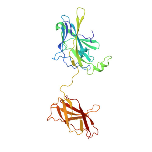
Structure of NFAT Bound to DNA as a Monomer

Stroud, J.C.Chen, L.

(2003) J Mol Biol 334: 1009-1022

  • DOI: https://doi.org/10.1016/j.jmb.2003.09.065
  • Primary Citation of Related Structures:  
    1OWR

  • PubMed Abstract: 

    The nuclear factor of activated T cells (NFAT) is a calcium-dependent transcription factor that cooperates with a myriad of partner transcription factors to regulate distinct transcription programs. Transcription activation by NFAT without the cooperation of co-stimulatory signals in lymphocytes can also impose a genetic program of anergy. Although the ternary NFAT1/Fos-Jun/DNA complex has been structurally characterized, how NFAT1 recognizes DNA in the absence of cooperative partners and how such a binary NFAT/DNA complex may lead to the assembly of distinct high-order NFAT transcription complexes are still poorly understood. We have determined the crystal structure of the entire Rel homology region (RHR) of human NFAT1 (NFATc2) bound to DNA as a monomer. We also present footprinting evidence that corroborates the protein-DNA contacts observed in the crystal structure. Our structural and biochemical studies reveal the mechanism by which the monomeric Rel protein NFAT recognizes its cognate DNA site. A remarkable feature of the binary NFAT/DNA complex is the conformational flexibility exhibited by NFAT1 in the four independent copies of the NFAT/DNA complex in the crystal structure, which may reflect a mechanism by which NFAT1 interacts with a variety of protein partners as it mediates disparate biological responses.


  • Organizational Affiliation

    Department of Chemistry and Biochemistry, University of Colorado at Boulder, Campus Box 215, Boulder, CO 80309-0437, USA.


Macromolecules

Find similar proteins by:  (by identity cutoff)  |  3D Structure
Entity ID: 3
MoleculeChains Sequence LengthOrganismDetailsImage
Nuclear factor of activated T-cells, cytoplasmic 2I [auth M],
J [auth N],
K [auth P],
L [auth Q]
284Homo sapiensMutation(s): 0 
Gene Names: NFATC2 OR NFAT1 OR NFATP
UniProt & NIH Common Fund Data Resources
Find proteins for Q13469 (Homo sapiens)
Explore Q13469 
Go to UniProtKB:  Q13469
PHAROS:  Q13469
GTEx:  ENSG00000101096 
Entity Groups  
Sequence Clusters30% Identity50% Identity70% Identity90% Identity95% Identity100% Identity
UniProt GroupQ13469
Sequence Annotations
Expand
  • Reference Sequence

Find similar nucleic acids by:  Sequence   |   3D Structure  

Entity ID: 1
MoleculeChains LengthOrganismImage
NFAT1 Monomeric Binding Site, Plus Strand
A, C, E, G
15N/A
Sequence Annotations
Expand
  • Reference Sequence

Find similar nucleic acids by:  Sequence   |   3D Structure  

Entity ID: 2
MoleculeChains LengthOrganismImage
NFAT1 Monomeric Binding Site, Minus Strand
B, D, F, H
15N/A
Sequence Annotations
Expand
  • Reference Sequence
Experimental Data & Validation

Experimental Data

  • Method: X-RAY DIFFRACTION
  • Resolution: 3.00 Å
  • R-Value Free: 0.273 
  • R-Value Work: 0.241 
  • Space Group: P 1 21 1
Unit Cell:
Length ( Å )Angle ( ˚ )
a = 100.646α = 90
b = 94.849β = 104.34
c = 112.788γ = 90
Software Package:
Software NamePurpose
DENZOdata reduction
SCALEPACKdata scaling
CNSrefinement
CNSphasing

Structure Validation

View Full Validation Report


Entry History 

Deposition Data

Revision History  (Full details and data files)

  • Version 1.0: 2004-02-10
    Type: Initial release
  • Version 1.1: 2008-04-29
    Changes: Version format compliance
  • Version 1.2: 2011-07-13
    Changes: Version format compliance
  • Version 1.3: 2023-08-16
    Changes: Data collection, Database references, Refinement description