Identification of Novel Binding Interactions in the Development of Potent, Selective 2-Naphthamidine Inhibitors of Urokinase. Synthesis, Structural Analysis, and SAR of N-Phenyl Amide 6-Substitution.
Wendt, M.D., Rockway, T.W., Geyer, A., McClellan, W., Weitzberg, M., Zhao, X., Mantei, R., Nienaber, V.L., Stewart, K., Klinghofer, V., Giranda, V.L.(2004) J Med Chem 47: 303-324
- PubMed: 14711304
- DOI: https://doi.org/10.1021/jm0300072
- Primary Citation of Related Structures:
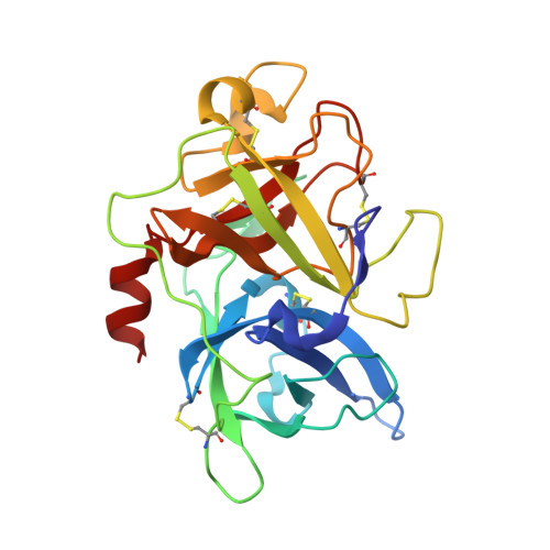
1OWD, 1OWE, 1OWH, 1OWI, 1OWJ, 1OWK - PubMed Abstract:
The preparation and assessment of biological activity of 6-substituted 2-naphthamidine inhibitors of the serine protease urokinase plasminogen activator (uPA, or urokinase) is described. 2-Naphthamidine was chosen as a starting point based on synthetic considerations and on modeling of substituent vectors. Phenyl amides at the 6-position were found to improve binding; replacement of the amide with other two-atom linkers proved ineffective. The phenyl group itself is situated near the S1' subsite; substitutions off of the phenyl group accessed S1' and other distant binding regions. Three new points of interaction were defined and explored through ring substitution. A solvent-exposed salt bridge with the Asp60A carboxylate was formed using a 4-alkylamino group, improving affinity to K(i) = 40 nM. Inhibitors also accessed two hydrophobic regions. One interaction is characterized by a tight hydrophobic fit made with a small dimple largely defined by His57 and His99; a weaker, less specific interaction involves alkyl groups reaching into the broad prime-side protein binding region near Val41 and the Cys42-Cys58 disulfide, displacing water molecules and leading to small gains in activity. Many inhibitors accessed two of these three regions. Affinities range as low as K(i) = 6 nM, and many compounds had K(i) < 100 nM, while moderate to excellent selectivity was gained versus four of five members of a panel of relevant serine proteases. Also, some selectivity against trypsin was generated via the interaction with Asp60A. X-ray structures of many of these compounds were used to inform our inhibitor design and to increase our understanding of key interactions. In combination with our exploration of 8-substitution patterns, we have identified a number of novel binding interactions for uPA inhibitors.
Organizational Affiliation:
Cancer Research and Structural Biology, Global Pharmaceutical R & D, Abbott Laboratories, 100 Abbott Park Road, Abbott Park, Illinois 60064-6101, USA. mike.d.wendt@abbott.com