1OVL

Crystal Structure of Nurr1 LBD


Experimental Data Snapshot

  • Method: X-RAY DIFFRACTION
  • Resolution: 2.20 Å
  • R-Value Free: 0.259 
  • R-Value Work: 0.217 
  • R-Value Observed: 0.231 

wwPDB Validation   3D Report Full Report


This is version 1.2 of the entry. See complete history


Literature

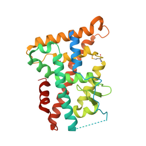
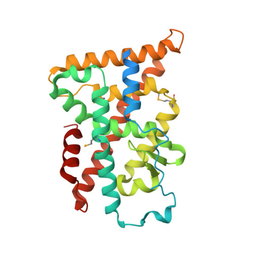
Structure and Function of Nurr1 identifies a Class of Ligand-Independent Nuclear Receptors

Wang, Z.Benoit, G.Liu, J.Prasad, S.Aarnisalo, P.Liu, X.Xu, H.Walker, N.Perlmann, T.

(2003) Nature 423: 555-560

  • DOI: https://doi.org/10.1038/nature01645
  • Primary Citation of Related Structures:  
    1OVL

  • PubMed Abstract: 

    Members of the nuclear receptor (NR) superfamily of transcription factors modulate gene transcription in response to small lipophilic molecules. Transcriptional activity is regulated by ligands binding to the carboxy-terminal ligand-binding domains (LBDs) of cognate NRs. A subgroup of NRs referred to as 'orphan receptors' lack identified ligands, however, raising issues about the function of their LBDs. Here we report the crystal structure of the LBD of the orphan receptor Nurr1 at 2.2 A resolution. The Nurr1 LBD adopts a canonical protein fold resembling that of agonist-bound, transcriptionally active LBDs in NRs, but the structure has two distinctive features. First, the Nurr1 LBD contains no cavity as a result of the tight packing of side chains from several bulky hydrophobic residues in the region normally occupied by ligands. Second, Nurr1 lacks a 'classical' binding site for coactivators. Despite these differences, the Nurr1 LBD can be regulated in mammalian cells. Notably, transcriptional activity is correlated with the Nurr1 LBD adopting a more stable conformation. Our findings highlight a unique structural class of NRs and define a model for ligand-independent NR function.


  • Organizational Affiliation

    Department of Structural Biology, Tularik Inc., 1120 Veterans Blvd., South San Francisco, California 94080, USA.


Macromolecules
Find similar proteins by:  (by identity cutoff)  |  3D Structure
Entity ID: 1
MoleculeChains Sequence LengthOrganismDetailsImage
Orphan nuclear receptor NURR1 (MSE 414, 496, 511)
A, D
271Homo sapiensMutation(s): 2 
Gene Names: NR4A2 OR NURR1 OR TINUR OR NOT
UniProt & NIH Common Fund Data Resources
Find proteins for P43354 (Homo sapiens)
Explore P43354 
Go to UniProtKB:  P43354
PHAROS:  P43354
GTEx:  ENSG00000153234 
Entity Groups  
Sequence Clusters30% Identity50% Identity70% Identity90% Identity95% Identity100% Identity
UniProt GroupP43354
Sequence Annotations
Expand
  • Reference Sequence
Find similar proteins by:  (by identity cutoff)  |  3D Structure
Entity ID: 2
MoleculeChains Sequence LengthOrganismDetailsImage
Orphan nuclear receptor NURR1 (MSE 496, 511)
B, C, E, F
271Homo sapiensMutation(s): 3 
Gene Names: NR4A2 OR NURR1 OR TINUR OR NOT
UniProt & NIH Common Fund Data Resources
Find proteins for P43354 (Homo sapiens)
Explore P43354 
Go to UniProtKB:  P43354
PHAROS:  P43354
GTEx:  ENSG00000153234 
Entity Groups  
Sequence Clusters30% Identity50% Identity70% Identity90% Identity95% Identity100% Identity
UniProt GroupP43354
Sequence Annotations
Expand
  • Reference Sequence
Small Molecules
Ligands 3 Unique
IDChains Name / Formula / InChI Key2D Diagram3D Interactions
IOD
Query on IOD

Download Ideal Coordinates CCD File 
P [auth B]IODIDE ION
I
XMBWDFGMSWQBCA-UHFFFAOYSA-M
BR
Query on BR

Download Ideal Coordinates CCD File 
AA [auth E]
BA [auth E]
CA [auth E]
DA [auth E]
EA [auth E]
AA [auth E],
BA [auth E],
CA [auth E],
DA [auth E],
EA [auth E],
GA [auth F],
H [auth A],
HA [auth F],
I [auth A],
J [auth A],
K [auth A],
O [auth B],
Q [auth C],
T [auth D],
U [auth D],
V [auth D],
W [auth D],
Y [auth E],
Z [auth E]
BROMIDE ION
Br
CPELXLSAUQHCOX-UHFFFAOYSA-M
K
Query on K

Download Ideal Coordinates CCD File 
FA [auth F]
G [auth A]
L [auth B]
M [auth B]
N [auth B]
FA [auth F],
G [auth A],
L [auth B],
M [auth B],
N [auth B],
R [auth D],
S [auth D],
X [auth E]
POTASSIUM ION
K
NPYPAHLBTDXSSS-UHFFFAOYSA-N
Modified Residues  1 Unique
IDChains TypeFormula2D DiagramParent
MSE
Query on MSE
A, D
L-PEPTIDE LINKINGC5 H11 N O2 SeMET
Experimental Data & Validation

Experimental Data

  • Method: X-RAY DIFFRACTION
  • Resolution: 2.20 Å
  • R-Value Free: 0.259 
  • R-Value Work: 0.217 
  • R-Value Observed: 0.231 
  • Space Group: P 31
Unit Cell:
Length ( Å )Angle ( ˚ )
a = 80.383α = 90
b = 80.383β = 90
c = 227.37γ = 120
Software Package:
Software NamePurpose
MOSFLMdata reduction
SCALAdata scaling
SHARPphasing
CNXrefinement
CCP4data scaling

Structure Validation

View Full Validation Report


Entry History 

Deposition Data

Revision History  (Full details and data files)

  • Version 1.0: 2003-06-03
    Type: Initial release
  • Version 1.1: 2008-04-29
    Changes: Version format compliance
  • Version 1.2: 2011-07-13
    Changes: Version format compliance