1OTR

Solution Structure of a CUE-Ubiquitin Complex


Experimental Data Snapshot

  • Method: SOLUTION NMR
  • Conformers Calculated: 80 
  • Conformers Submitted: 20 
  • Selection Criteria: The submitted conformer models are the 20 structures with the lowest restraint energies, restraint violations, and RMS deviations from ideal covalent geometry 

wwPDB Validation   3D Report Full Report


This is version 1.3 of the entry. See complete history


Literature

Solution Structure of a CUE-Ubiquitin Complex Reveals a Conserved Mode of Ubiquitin Binding

Kang, R.S.Daniels, C.M.Francis, S.A.Shih, S.C.Salerno, W.J.Hicke, L.Radhakrishnan, I.

(2003) Cell 113: 621-630

  • DOI: https://doi.org/10.1016/s0092-8674(03)00362-3
  • Primary Citation of Related Structures:  
    1OTR

  • PubMed Abstract: 

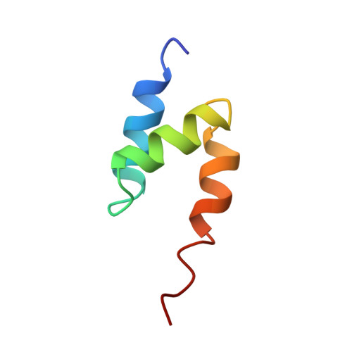
    Monoubiquitination serves as a regulatory signal in a variety of cellular processes. Monoubiquitin signals are transmitted by binding to a small but rapidly expanding class of ubiquitin binding motifs. Several of these motifs, including the CUE domain, also promote intramolecular monoubiquitination. The solution structure of a CUE domain of the yeast Cue2 protein in complex with ubiquitin reveals intermolecular interactions involving conserved hydrophobic surfaces, including the Leu8-Ile44-Val70 patch on ubiquitin. The contact surface extends beyond this patch and encompasses Lys48, a site of polyubiquitin chain formation. This suggests an occlusion mechanism for inhibiting polyubiquitin chain formation during monoubiquitin signaling. The CUE domain shares a similar overall architecture with the UBA domain, which also contains a conserved hydrophobic patch. Comparative modeling suggests that the UBA domain interacts analogously with ubiquitin. The structure of the CUE-ubiquitin complex may thus serve as a paradigm for ubiquitin recognition and signaling by ubiquitin binding proteins.


  • Organizational Affiliation

    Department of Biochemistry, Molecular Biology, and Cell Biology, Northwestern University, Evanston, IL 60208, USA.


Macromolecules
Find similar proteins by:  (by identity cutoff)  |  3D Structure
Entity ID: 1
MoleculeChains Sequence LengthOrganismDetailsImage
protein Cue249Saccharomyces cerevisiaeMutation(s): 0 
Gene Names: CUE2
UniProt
Find proteins for P36075 (Saccharomyces cerevisiae (strain ATCC 204508 / S288c))
Explore P36075 
Go to UniProtKB:  P36075
Entity Groups  
Sequence Clusters30% Identity50% Identity70% Identity90% Identity95% Identity100% Identity
UniProt GroupP36075
Sequence Annotations
Expand
  • Reference Sequence
Find similar proteins by:  (by identity cutoff)  |  3D Structure
Entity ID: 2
MoleculeChains Sequence LengthOrganismDetailsImage
Ubiquitin76Saccharomyces cerevisiaeMutation(s): 0 
Gene Names: UBI1
UniProt
Find proteins for P0CG63 (Saccharomyces cerevisiae (strain ATCC 204508 / S288c))
Explore P0CG63 
Go to UniProtKB:  P0CG63
Entity Groups  
Sequence Clusters30% Identity50% Identity70% Identity90% Identity95% Identity100% Identity
UniProt GroupP0CG63
Sequence Annotations
Expand
  • Reference Sequence
Experimental Data & Validation

Experimental Data

  • Method: SOLUTION NMR
  • Conformers Calculated: 80 
  • Conformers Submitted: 20 
  • Selection Criteria: The submitted conformer models are the 20 structures with the lowest restraint energies, restraint violations, and RMS deviations from ideal covalent geometry 

Structure Validation

View Full Validation Report


Entry History 

Deposition Data

Revision History  (Full details and data files)

  • Version 1.0: 2003-06-24
    Type: Initial release
  • Version 1.1: 2008-04-29
    Changes: Version format compliance
  • Version 1.2: 2011-07-13
    Changes: Version format compliance
  • Version 1.3: 2022-02-23
    Changes: Data collection, Database references, Derived calculations