1.85-A resolution crystal structure of human ornithine transcarbamoylase complexed with N-phosphonacetyl-L-ornithine. Catalytic mechanism and correlation with inherited deficiency.
Shi, D., Morizono, H., Ha, Y., Aoyagi, M., Tuchman, M., Allewell, N.M.(1998) J Biol Chem 273: 34247-34254
- PubMed: 9852088
- DOI: https://doi.org/10.1074/jbc.273.51.34247
- Primary Citation of Related Structures:
1OTH - PubMed Abstract:
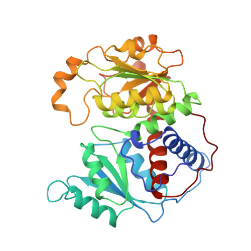
The crystal structure of human ornithine transcarbamoylase complexed with the bisubstrate analog N-phosphonacetyl-L-ornithine has been solved at 1.85-A resolution by molecular replacement. Deleterious mutations produce clinical hyperammonia that, if untreated, results in neurological symptoms or death (ornithine transcarbamylase deficiency). The holoenzyme is trimeric, and as in other transcarbamoylases, each subunit contains an N-terminal domain that binds carbamoyl phosphate and a C-terminal domain that binds L-ornithine. The active site is located in the cleft between domains and contains additional residues from an adjacent subunit. Binding of N-phosphonacetyl-L-ornithine promotes domain closure. The resolution of the structure enables the role of active site residues in the catalytic mechanism to be critically examined. The side chain of Cys-303 is positioned so as to be able to interact with the delta-amino group of L-ornithine which attacks the carbonyl carbon of carbamoyl phosphate in the enzyme-catalyzed reaction. This sulfhydryl group forms a charge relay system with Asp-263 and the alpha-amino group of L-ornithine, instead of with His-302 and Glu-310, as previously proposed. In common with other ureotelic ornithine transcarbamoylases, the human enzyme lacks a loop of approximately 20 residues between helix H10 and beta-strand B10 which is present in prokaryotic ornithine transcarbamoylases but has a C-terminal extension of 10 residues that interacts with the body of the protein but is exposed. The sequence of this C-terminal extension is homologous to an interhelical loop found in several membrane proteins, including mitochondrial transport proteins, suggesting a possible mode of interaction with the inner mitochondrial membrane.
Organizational Affiliation:
Department of Biochemistry, University of Minnesota, St. Paul, Minnesota 55108, USA.