1OT3

Crystal structure of Drosophila deoxyribonucleotide kinase complexed with the substrate deoxythymidine


Experimental Data Snapshot

  • Method: X-RAY DIFFRACTION
  • Resolution: 2.50 Å
  • R-Value Free: 0.276 
  • R-Value Work: 0.227 

wwPDB Validation   3D Report Full Report


Ligand Structure Quality Assessment 


This is version 1.4 of the entry. See complete history


Literature

Structural Basis for Feedback Inhibition of the Deoxyribonucleoside Salvage Pathway: Studies of the Drosophila Deoxyribonucleoside Kinase.

Mikkelsen, N.E.Johansson, K.Karlsson, A.Knecht, W.Andersen, G.Piskur, J.Munch-Petersen, B.Eklund, H.

(2003) Biochemistry 42: 5706-5712

  • DOI: https://doi.org/10.1021/bi0340043
  • Primary Citation of Related Structures:  
    1OE0, 1OT3

  • PubMed Abstract: 

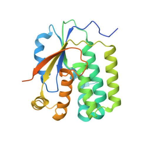
    Deoxyribonucleoside kinases are feedback inhibited by the final products of the salvage pathway, the deoxyribonucleoside triphosphates. In the present study, the mechanism of feedback inhibition is presented based on the crystal structure of a complex between the fruit fly deoxyribonucleoside kinase and its feedback inhibitor deoxythymidine triphosphate. The inhibitor was found to be bound as a bisubstrate inhibitor with its nucleoside part in the nucleoside binding site and with its phosphate groups partially occupying the phosphate donor site. The overall structure of the enzyme--inhibitor complex is very similar to the enzyme--substrate complexes with deoxythymidine and deoxycytidine, except for a conformational change within a region otherwise directly involved in catalysis. This conformational change involves a magnesium ion, which is coordinated in the inhibitor complex to the phosphates and to the primary base, Glu52, that normally is positioned close to the 5'-OH of the substrate deoxyribose.


  • Organizational Affiliation

    Department of Molecular Biology, Swedish University of Agricultural Sciences, Box 590, Biomedical Center, S-751 24 Uppsala, Sweden.


Macromolecules
Find similar proteins by:  (by identity cutoff)  |  3D Structure
Entity ID: 1
MoleculeChains Sequence LengthOrganismDetailsImage
Deoxyribonucleoside Kinase
A, B, C, D, E
A, B, C, D, E, F, G, H
250Drosophila melanogasterMutation(s): 0 
EC: 2.7.1.145
UniProt
Find proteins for Q9XZT6 (Drosophila melanogaster)
Explore Q9XZT6 
Go to UniProtKB:  Q9XZT6
Entity Groups  
Sequence Clusters30% Identity50% Identity70% Identity90% Identity95% Identity100% Identity
UniProt GroupQ9XZT6
Sequence Annotations
Expand
  • Reference Sequence
Small Molecules
Ligands 2 Unique
IDChains Name / Formula / InChI Key2D Diagram3D Interactions
THM
Query on THM

Download Ideal Coordinates CCD File 
K [auth A]
M [auth B]
O [auth C]
Q [auth D]
S [auth E]
K [auth A],
M [auth B],
O [auth C],
Q [auth D],
S [auth E],
U [auth F],
W [auth G],
Y [auth H]
THYMIDINE
C10 H14 N2 O5
IQFYYKKMVGJFEH-XLPZGREQSA-N
SO4
Query on SO4

Download Ideal Coordinates CCD File 
I [auth A]
J [auth A]
L [auth B]
N [auth C]
P [auth D]
I [auth A],
J [auth A],
L [auth B],
N [auth C],
P [auth D],
R [auth E],
T [auth F],
V [auth G],
X [auth H]
SULFATE ION
O4 S
QAOWNCQODCNURD-UHFFFAOYSA-L
Experimental Data & Validation

Experimental Data

  • Method: X-RAY DIFFRACTION
  • Resolution: 2.50 Å
  • R-Value Free: 0.276 
  • R-Value Work: 0.227 
  • Space Group: P 1 21 1
Unit Cell:
Length ( Å )Angle ( ˚ )
a = 70.379α = 90
b = 71.158β = 90.59
c = 226.272γ = 90
Software Package:
Software NamePurpose
MOSFLMdata reduction
SCALAdata scaling
MOLREPphasing
CNSrefinement
CCP4data scaling

Structure Validation

View Full Validation Report


Ligand Structure Quality Assessment 


Entry History 

Deposition Data

Revision History  (Full details and data files)

  • Version 1.0: 2003-05-27
    Type: Initial release
  • Version 1.1: 2008-04-29
    Changes: Version format compliance
  • Version 1.2: 2011-07-13
    Changes: Version format compliance
  • Version 1.3: 2014-05-28
    Changes: Other
  • Version 1.4: 2023-10-25
    Changes: Data collection, Database references, Derived calculations, Refinement description