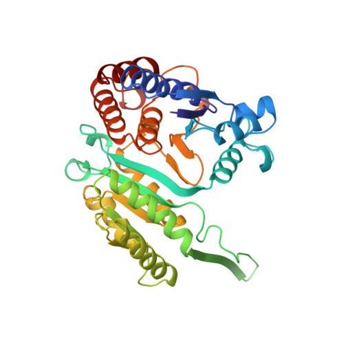
A mutation at the interface between domains causes rearrangement of domains in 3-isopropylmalate dehydrogenase.
Qu, C., Akanuma, S., Moriyama, H., Tanaka, N., Oshima, T.(1997) Protein Eng 10: 45-52
- PubMed: 9051733
- DOI: https://doi.org/10.1093/protein/10.1.45
- Primary Citation of Related Structures:
1OSI, 1OSJ - PubMed Abstract:
The structure of a thermostable Ala172Leu mutant, designated A172L, of 3-isopropylmalate dehydrogenase from Thermus thermophilus was determined. The crystal belongs to space group P2(1), with cell parameters a = 55.5 A, b = 88.1 A, c = 72.0 A and beta = 100.9 degrees. There is one dimer in each asymmetric unit. The final R factor is 17.8% with 69 water molecules at 2.35 A resolution. The mutation is located at the interface between domains and the C alpha trace of the mutant structure deviates from that of the native structure by as much as 1.7 A, while the structure of each domain barely changes. The mutant enzyme has a more closed conformation compared with the wild-type enzyme as a result of the replacement of Ala with Leu at residue 172. These structural variations were found independent of the crystal packing, because the structure of wild type was the same in crystals obtained in different precipitants. The hinge regions for the movement of domains are located around the active cleft of the enzyme, an observation that implies that the mobility of domains around the hinge is indispensable for the activity of the enzyme. The larger side chain at the mutated site contributed to the thermostability of the mutant protein by enhancing the local packing of side chains, and also by shifting the backbone of the opposing domain.
Organizational Affiliation:
Department of Life Science, Faculty of Bioscience and Biotechnology, Tokyo Institute of Technology, Yokohama, Japan.