1OSC

Crystal structure of rat CUTA1 at 2.15 A resolution


Experimental Data Snapshot

  • Method: X-RAY DIFFRACTION
  • Resolution: 2.15 Å
  • R-Value Free: 0.260 
  • R-Value Work: 0.189 
  • R-Value Observed: 0.195 

wwPDB Validation   3D Report Full Report


This is version 1.4 of the entry. See complete history


Literature

The Evolutionarily Conserved Trimeric Structure of CutA1 Proteins Suggests a Role in Signal Transduction

Arnesano, F.Banci, L.Benvenuti, M.Bertini, I.Calderone, V.Mangani, S.Viezzoli, M.S.

(2003) J Biol Chem 278: 45999-46006

  • DOI: https://doi.org/10.1074/jbc.M304398200
  • Primary Citation of Related Structures:  
    1NAQ, 1OSC

  • PubMed Abstract: 

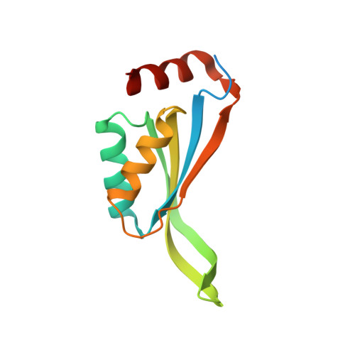
    CutA1 are a protein family present in bacteria, plants, and animals, including humans. Escherichia coli CutA1 is involved in copper tolerance, whereas mammalian proteins are implicated in the anchoring of acetylcholinesterase in neuronal cell membranes. The x-ray structures of CutA1 from E. coli and rat were determined. Both proteins are trimeric in the crystals and in solution through an inter-subunit beta-sheet formation. Each subunit consists of a ferredoxin-like (beta1alpha1beta2beta3alpha2beta4) fold with an additional strand (beta5), a C-terminal helix (alpha3), and an unusual extended beta-hairpin involving strands beta2 and beta3. The bacterial CutA1 is able to bind copper(II) in vitro through His2Cys coordination in a type II water-accessible site, whereas the rat protein precipitates in the presence of copper(II). The evolutionarily conserved trimeric assembly of CutA1 is reminiscent of the architecture of PII signal transduction proteins. This similarity suggests an intriguing role of CutA1 proteins in signal transduction through allosteric communications between subunits.


  • Organizational Affiliation

    Magnetic Resonance Center (CERM) and Department of Chemistry, University of Florence, Via Luigi Sacconi 6, 50019, Sesto Fiorentino, Florence, Italy.


Macromolecules
Find similar proteins by:  (by identity cutoff)  |  3D Structure
Entity ID: 1
MoleculeChains Sequence LengthOrganismDetailsImage
similar to divalent cation tolerant protein CUTA
A, B, C, D, E
A, B, C, D, E, F
126Rattus norvegicusMutation(s): 0 
Gene Names: CutACutA1
UniProt
Find proteins for Q6MGD0 (Rattus norvegicus)
Explore Q6MGD0 
Go to UniProtKB:  Q6MGD0
Entity Groups  
Sequence Clusters30% Identity50% Identity70% Identity90% Identity95% Identity100% Identity
UniProt GroupQ6MGD0
Sequence Annotations
Expand
  • Reference Sequence
Experimental Data & Validation

Experimental Data

  • Method: X-RAY DIFFRACTION
  • Resolution: 2.15 Å
  • R-Value Free: 0.260 
  • R-Value Work: 0.189 
  • R-Value Observed: 0.195 
  • Space Group: P 21 21 21
Unit Cell:
Length ( Å )Angle ( ˚ )
a = 70.387α = 90
b = 88.287β = 90
c = 125.853γ = 90
Software Package:
Software NamePurpose
REFMACrefinement
MOSFLMdata reduction
CCP4data scaling
MOLREPphasing

Structure Validation

View Full Validation Report


Entry History 

Revision History  (Full details and data files)

  • Version 1.0: 2003-11-25
    Type: Initial release
  • Version 1.1: 2008-04-29
    Changes: Version format compliance
  • Version 1.2: 2011-07-13
    Changes: Version format compliance
  • Version 1.3: 2018-01-24
    Changes: Database references
  • Version 1.4: 2023-08-16
    Changes: Data collection, Database references, Refinement description