Structural basis of fosmidomycin action revealed by the complex with 2-C-methyl-D-erythritol 4-phosphate synthase (IspC). Implications for the catalytic mechanism and anti-malaria drug development.
Steinbacher, S., Kaiser, J., Eisenreich, W., Huber, R., Bacher, A., Rohdich, F.(2003) J Biol Chem 278: 18401-18407
- PubMed: 12621040
- DOI: https://doi.org/10.1074/jbc.M300993200
- Primary Citation of Related Structures:
1ONN, 1ONO, 1ONP - PubMed Abstract:
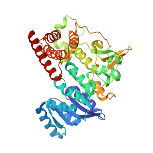
2-C-Methyl-d-erythritol 4-phosphate synthase (IspC) is the first enzyme committed to isoprenoid biosynthesis in the methylerythritol phosphate pathway, which represents an alternative route to the classical mevalonate pathway. As it is present in many pathogens and plants, but not in man, this pathway has attracted considerable interest as a target for novel antibiotics and herbicides. Fosmidomycin represents a specific high-affinity inhibitor of IspC. Very recently, its anti-malaria activity in man has been demonstrated in clinical trials. Here, we present the crystal structure of Escherichia coli IspC in complex with manganese and fosmidomycin at 2.5 A resolution. The (N-formyl-N-hydroxy)amino group provides two oxygen ligands to manganese that is present in a distorted octahedral coordination, whereas the phosphonate group is anchored in a specific pocket by numerous hydrogen bonds. Both sites are connected by a spacer of three methylene groups. The substrate molecule, 1-d-deoxyxylulose 5-phosphate, can be superimposed onto fosmidomycin, explaining the stereochemical course of the reaction.
Organizational Affiliation:
Max-Planck-Institut für Biochemie, Abteilung für Strukturforschung, Am Klopferspitz 18a, D-82152 Martinsried, Germany. steinbac@caltech.edu