1ONF

Crystal structure of Plasmodium falciparum Glutathione reductase


Experimental Data Snapshot

  • Method: X-RAY DIFFRACTION
  • Resolution: 2.60 Å
  • R-Value Free: 0.297 
  • R-Value Work: 0.256 
  • R-Value Observed: 0.260 

wwPDB Validation   3D Report Full Report


Ligand Structure Quality Assessment 


This is version 1.3 of the entry. See complete history


Literature

Glutathione reductase of the malarial parasite Plasmodium falciparum: Crystal structure and inhibitor development

Sarma, G.N.Savvides, S.N.Becker, K.Schirmer, M.Schirmer, R.H.Karplus, P.A.

(2003) J Mol Biol 328: 893-907

  • DOI: https://doi.org/10.1016/s0022-2836(03)00347-4
  • Primary Citation of Related Structures:  
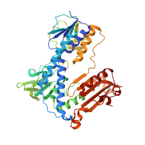
    1ONF

  • PubMed Abstract: 

    The malarial parasite Plasmodium falciparum is known to be sensitive to oxidative stress, and thus the antioxidant enzyme glutathione reductase (GR; NADPH+GSSG+H(+) <==> NADP(+)+2 GSH) has become an attractive drug target for antimalarial drug development. Here, we report the 2.6A resolution crystal structure of P.falciparum GR. The homodimeric flavoenzyme is compared to the related human GR with focus on structural aspects relevant for drug design. The most pronounced differences between the two enzymes concern the shape and electrostatics of a large (450A(3)) cavity at the dimer interface. This cavity binds numerous non-competitive inhibitors and is a target for selective drug design. A 34-residue insertion specific for the GRs of malarial parasites shows no density, implying that it is disordered. The precise location of this insertion along the sequence allows us to explain the deleterious effects of a mutant in this region and suggests new functional studies. To complement the structural comparisons, we report the relative susceptibility of human and plasmodial GRs to a series of tricyclic inhibitors as well as to peptides designed to interfere with protein folding and dimerization. Enzyme-kinetic studies on GRs from chloroquine-resistant and chloroquine-sensitive parasite strains were performed and indicate that the structure reported here represents GR of P.falciparum strains in general and thus is a highly relevant target for drug development.


  • Organizational Affiliation

    Department of Biochemistry and Biophysics, Oregon State University, Corvallis, OR 97331-7305, USA.


Macromolecules
Find similar proteins by:  (by identity cutoff)  |  3D Structure
Entity ID: 1
MoleculeChains Sequence LengthOrganismDetailsImage
Glutathione reductase500Plasmodium falciparumMutation(s): 0 
Gene Names: GR2
EC: 1.8.1.7
UniProt
Find proteins for Q94655 (Plasmodium falciparum (isolate K1 / Thailand))
Explore Q94655 
Go to UniProtKB:  Q94655
Entity Groups  
Sequence Clusters30% Identity50% Identity70% Identity90% Identity95% Identity100% Identity
UniProt GroupQ94655
Sequence Annotations
Expand
  • Reference Sequence
Small Molecules
Ligands 1 Unique
IDChains Name / Formula / InChI Key2D Diagram3D Interactions
FAD
Query on FAD

Download Ideal Coordinates CCD File 
B [auth A]FLAVIN-ADENINE DINUCLEOTIDE
C27 H33 N9 O15 P2
VWWQXMAJTJZDQX-UYBVJOGSSA-N
Experimental Data & Validation

Experimental Data

  • Method: X-RAY DIFFRACTION
  • Resolution: 2.60 Å
  • R-Value Free: 0.297 
  • R-Value Work: 0.256 
  • R-Value Observed: 0.260 
  • Space Group: P 41 2 2
Unit Cell:
Length ( Å )Angle ( ˚ )
a = 90.64α = 90
b = 90.64β = 90
c = 123.32γ = 90
Software Package:
Software NamePurpose
REFMACrefinement
DENZOdata reduction
SCALEPACKdata scaling
CNSphasing

Structure Validation

View Full Validation Report


Ligand Structure Quality Assessment 


Entry History 

Deposition Data

Revision History  (Full details and data files)

  • Version 1.0: 2003-05-06
    Type: Initial release
  • Version 1.1: 2008-04-29
    Changes: Version format compliance
  • Version 1.2: 2011-07-13
    Changes: Derived calculations, Version format compliance
  • Version 1.3: 2023-08-16
    Changes: Data collection, Database references, Derived calculations, Refinement description